 Movie
Movie Controller

[English] 日本語
 Yorodumi
- PDB-3f52: Crystal structure of the clp gene regulator ClgR from C. glutamicum -
+ Open data

- Basic information
| Entry | Database: PDB / ID: 3f52 | ||||||
|---|---|---|---|---|---|---|---|
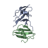
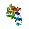
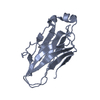
| Title | Crystal structure of the clp gene regulator ClgR from C. glutamicum | ||||||
 Components Components | clp gene regulator (ClgR) | ||||||
 Keywords Keywords | TRANSCRIPTION ACTIVATOR / gene regulator / helix-turn-helix motif / transcriptional activator / human pathogen | ||||||
| Function / homology |  Function and homology information Function and homology informationDNA-binding transcription activator activity / protein-DNA complex / transcription cis-regulatory region binding / positive regulation of DNA-templated transcription / DNA-templated transcription / cytosol Similarity search - Function | ||||||
| Biological species |  Corynebacterium glutamicum (bacteria) Corynebacterium glutamicum (bacteria) | ||||||
| Method |  X-RAY DIFFRACTION / SYNCHROTRON / MOLECULAR REPLACEMENT / Resolution: 1.75 Å X-RAY DIFFRACTION / SYNCHROTRON / MOLECULAR REPLACEMENT / Resolution: 1.75 Å | ||||||
 Authors Authors | Russo, S. / Schweitzer, J.E. / Polen, T. / Bott, M. / Pohl, E. | ||||||
 Citation Citation | Journal: J.Biol.Chem. / Year: 2009 Title: Crystal structure of the caseinolytic protease gene regulator, a transcriptional activator in actinomycetes Authors: Russo, S. / Schweitzer, J.E. / Polen, T. / Bott, M. / Pohl, E. | ||||||
| History |
|
- Structure visualization
| Structure viewer | Molecule: MolmilJmol/JSmol |
|---|
- Downloads & links
-Download
| PDBx/mmCIF format | 3f52.cif.gz | 46.5 KB | Display | PDBx/mmCIF format |
|---|---|---|---|---|
| PDB format | pdb3f52.ent.gz | 32 KB | Display | PDB format |
| PDBx/mmJSON format | 3f52.json.gz | Tree view | PDBx/mmJSON format | |
| Others |  Other downloads Other downloads |
-Validation report
| Arichive directory | https://data.pdbj.org/pub/pdb/validation_reports/f5/3f52ftp://data.pdbj.org/pub/pdb/validation_reports/f5/3f52 | HTTPS FTP |
|---|
-Related structure data
| Related structure data | 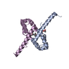 3f51SC S: Starting model for refinement C: citing same article ( |
|---|---|
| Similar structure data |


-Links
 PDBj
PDBj


- Assembly
| Deposited unit | 
| ||||||||
|---|---|---|---|---|---|---|---|---|---|
| 1 |
| ||||||||
| Unit cell |
|
-Components
| #1: Protein | Mass: 12647.396 Da / Num. of mol.: 2 / Fragment: clgr_c Source method: isolated from a genetically manipulated source Source: (gene. exp.) Corynebacterium glutamicum (bacteria) / Gene: cg2152, Cgl1962 / Plasmid: pEKEx1 / Production host: #2: Chemical | ChemComp-GOL /   Mass: 92.094 Da / Num. of mol.: 5 / Source method: obtained synthetically / Formula: C3H8O3 Mass: 92.094 Da / Num. of mol.: 5 / Source method: obtained synthetically / Formula: C3H8O3#3: Water | ChemComp-HOH / |  Mass: 18.015 Da / Num. of mol.: 156 / Source method: isolated from a natural source / Formula: H2O Mass: 18.015 Da / Num. of mol.: 156 / Source method: isolated from a natural source / Formula: H2O |
|---|
-Experimental details
-Experiment
| Experiment | Method: X-RAY DIFFRACTION / Number of used crystals: 1 |
|---|
- Sample preparation
| Crystal | Density Matthews: 1.94 Å3/Da / Density % sol: 36.7 % |
|---|---|
| Crystal grow | Temperature: 298 K / Method: vapor diffusion, sitting drop / pH: 7.5 Details: 0.085 M HEPES, 8.5% PEG 8000, 15% glycerol, pH 7.5, VAPOR DIFFUSION, SITTING DROP, temperature 298K |
-Data collection
| Diffraction | Mean temperature: 100 K |
|---|---|
| Diffraction source | Source: SYNCHROTRON / Site: SLS  / Beamline: X10SA / Wavelength: 1.0685 Å / Beamline: X10SA / Wavelength: 1.0685 Å |
| Detector | Type: MARMOSAIC 225 mm CCD / Detector: CCD / Date: Apr 16, 2008 |
| Radiation | Monochromator: sagitally focused Si(111), bending mirror for vertical focusing, spot size 20x50um Protocol: SINGLE WAVELENGTH / Monochromatic (M) / Laue (L): M / Scattering type: x-ray |
| Radiation wavelength | Wavelength: 1.0685 Å / Relative weight: 1 |
| Reflection | Resolution: 1.75→42 Å / Num. all: 20944 / Num. obs: 20938 / % possible obs: 100 % / Observed criterion σ(F): 1.7 / Observed criterion σ(I): 3 / Redundancy: 10 % / Rsym value: 0.057 / Net I/σ(I): 24.56 |
| Reflection shell | Resolution: 1.75→1.85 Å / Redundancy: 8.5 % / Mean I/σ(I) obs: 2.56 / Num. unique all: 3143 / Rsym value: 0.799 / % possible all: 100 |
- Processing
| Software |
| ||||||||||||||||||||||||||||||||||||||||||||||||||||||||||||||||||||||||||||||||||||||||||
|---|---|---|---|---|---|---|---|---|---|---|---|---|---|---|---|---|---|---|---|---|---|---|---|---|---|---|---|---|---|---|---|---|---|---|---|---|---|---|---|---|---|---|---|---|---|---|---|---|---|---|---|---|---|---|---|---|---|---|---|---|---|---|---|---|---|---|---|---|---|---|---|---|---|---|---|---|---|---|---|---|---|---|---|---|---|---|---|---|---|---|---|
| Refinement | Method to determine structure: MOLECULAR REPLACEMENT Starting model: PDB entry 3F51 Resolution: 1.75→41.96 Å / Cor.coef. Fo:Fc: 0.962 / Cor.coef. Fo:Fc free: 0.957 / SU B: 4.273 / SU ML: 0.069 / TLS residual ADP flag: LIKELY RESIDUAL / Cross valid method: THROUGHOUT / σ(F): 1.7 / σ(I): 3 / ESU R: 0.098 / ESU R Free: 0.097 / Stereochemistry target values: MAXIMUM LIKELIHOOD / Details: HYDROGENS HAVE BEEN ADDED IN THE RIDING POSITIONS
| ||||||||||||||||||||||||||||||||||||||||||||||||||||||||||||||||||||||||||||||||||||||||||
| Solvent computation | Ion probe radii: 0.8 Å / Shrinkage radii: 0.8 Å / VDW probe radii: 1.2 Å / Solvent model: MASK | ||||||||||||||||||||||||||||||||||||||||||||||||||||||||||||||||||||||||||||||||||||||||||
| Displacement parameters | Biso mean: 33.788 Å2
| ||||||||||||||||||||||||||||||||||||||||||||||||||||||||||||||||||||||||||||||||||||||||||
| Refinement step | Cycle: LAST / Resolution: 1.75→41.96 Å
| ||||||||||||||||||||||||||||||||||||||||||||||||||||||||||||||||||||||||||||||||||||||||||
| Refine LS restraints |
| ||||||||||||||||||||||||||||||||||||||||||||||||||||||||||||||||||||||||||||||||||||||||||
| LS refinement shell | Resolution: 1.75→1.795 Å / Total num. of bins used: 20
| ||||||||||||||||||||||||||||||||||||||||||||||||||||||||||||||||||||||||||||||||||||||||||
| Refinement TLS params. | Method: refined / Refine-ID: X-RAY DIFFRACTION
| ||||||||||||||||||||||||||||||||||||||||||||||||||||||||||||||||||||||||||||||||||||||||||
| Refinement TLS group |
|
