 Movie
Movie Controller


+ Open data

- Basic information
| Entry | Database: PDB / ID: 5fud | ||||||
|---|---|---|---|---|---|---|---|
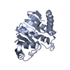
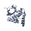
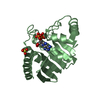
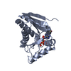
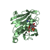
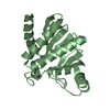
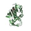
| Title | Oceanobacillus iheyensis macrodomain with MES bound | ||||||
 Components Components | O-ACETYL-ADP-RIBOSE DEACETYLASE | ||||||
 Keywords Keywords | HYDROLASE / BACTERIAL MACRODOMAIN / ADP-RIBOSE / DEACETYLASE | ||||||
| Function / homology |  Function and homology information Function and homology information | ||||||
| Biological species |  OCEANOBACILLUS IHEYENSIS (bacteria) OCEANOBACILLUS IHEYENSIS (bacteria) | ||||||
| Method |  X-RAY DIFFRACTION / SYNCHROTRON / MOLECULAR REPLACEMENT / Resolution: 1.9 Å X-RAY DIFFRACTION / SYNCHROTRON / MOLECULAR REPLACEMENT / Resolution: 1.9 Å | ||||||
 Authors Authors | Gil-Ortiz, F. / Zapata-Perez, R. / Martinez, A.B. / Juanhuix, J. / Sanchez-Ferrer, A. | ||||||
 Citation Citation | Journal: Open Biol / Year: 2017 Title: Structural and functional analysis ofOceanobacillus iheyensismacrodomain reveals a network of waters involved in substrate binding and catalysis. Authors: Zapata-Perez, R. / Gil-Ortiz, F. / Martinez-Monino, A.B. / Garcia-Saura, A.G. / Juanhuix, J. / Sanchez-Ferrer, A. | ||||||
| History |
|
- Structure visualization
| Structure viewer | Molecule: MolmilJmol/JSmol |
|---|
- Downloads & links
-Download
| PDBx/mmCIF format | 5fud.cif.gz | 89.4 KB | Display | PDBx/mmCIF format |
|---|---|---|---|---|
| PDB format | pdb5fud.ent.gz | 67 KB | Display | PDB format |
| PDBx/mmJSON format | 5fud.json.gz | Tree view | PDBx/mmJSON format | |
| Others |  Other downloads Other downloads |
-Validation report
| Arichive directory | https://data.pdbj.org/pub/pdb/validation_reports/fu/5fudftp://data.pdbj.org/pub/pdb/validation_reports/fu/5fud | HTTPS FTP |
|---|
-Related structure data
| Related structure data |  5l9kC  5l9qC  5lauC  5lbpC  5lccC 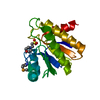 1spvS S: Starting model for refinement C: citing same article ( |
|---|---|
| Similar structure data |


-Links
 PDBj
PDBj- Assembly
| Deposited unit | 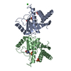
| ||||||||||||||||||||||||||||||||||||||||||
|---|---|---|---|---|---|---|---|---|---|---|---|---|---|---|---|---|---|---|---|---|---|---|---|---|---|---|---|---|---|---|---|---|---|---|---|---|---|---|---|---|---|---|---|
| 1 | 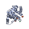
| ||||||||||||||||||||||||||||||||||||||||||
| 2 | 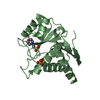
| ||||||||||||||||||||||||||||||||||||||||||
| Unit cell |
| ||||||||||||||||||||||||||||||||||||||||||
| Noncrystallographic symmetry (NCS) | NCS domain:
NCS domain segments: Component-ID: 1 / Ens-ID: 1 / End auth comp-ID: ILE / End label comp-ID: ILE / Refine code: 6
NCS oper:
|
-Components
| #1: Protein | Mass: 22801.857 Da / Num. of mol.: 2 Source method: isolated from a genetically manipulated source Source: (gene. exp.) OCEANOBACILLUS IHEYENSIS (bacteria) / Strain: HTE831 / Production host: References: UniProt: Q8EP31, Hydrolases; Glycosylases; Hydrolysing N-glycosyl compounds, Hydrolases; Acting on carbon-nitrogen bonds, other than peptide bonds; In linear amides #2: Chemical |   Mass: 96.063 Da / Num. of mol.: 2 / Source method: obtained synthetically / Formula: SO4 Mass: 96.063 Da / Num. of mol.: 2 / Source method: obtained synthetically / Formula: SO4#3: Chemical | ChemComp-CL / |   Mass: 35.453 Da / Num. of mol.: 1 / Source method: obtained synthetically / Formula: Cl Mass: 35.453 Da / Num. of mol.: 1 / Source method: obtained synthetically / Formula: Cl#4: Chemical |   Mass: 195.237 Da / Num. of mol.: 2 / Source method: obtained synthetically / Formula: C6H13NO4S / Comment: pH buffer*YM Mass: 195.237 Da / Num. of mol.: 2 / Source method: obtained synthetically / Formula: C6H13NO4S / Comment: pH buffer*YM#5: Water | ChemComp-HOH / |  Mass: 18.015 Da / Num. of mol.: 218 / Source method: isolated from a natural source / Formula: H2O Mass: 18.015 Da / Num. of mol.: 218 / Source method: isolated from a natural source / Formula: H2O |
|---|
-Experimental details
-Experiment
| Experiment | Method: X-RAY DIFFRACTION / Number of used crystals: 1 |
|---|
- Sample preparation
| Crystal | Density Matthews: 2.74 Å3/Da / Density % sol: 55 % / Description: NONE |
|---|---|
| Crystal grow | pH: 6.5 Details: 0.2 M AMMONIUM SULPHATE, 0.1 M MES, PH 6.5, 30% (W/V) PEG [POLY(ETHYLENE GLYCOL)] MME 5000 |
-Data collection
| Diffraction | Mean temperature: 100 K | |||||||||||||||
|---|---|---|---|---|---|---|---|---|---|---|---|---|---|---|---|---|
| Diffraction source | Source: SYNCHROTRON / Site: ALBA  / Beamline: XALOC / Wavelength: 0.97944 / Beamline: XALOC / Wavelength: 0.97944 | |||||||||||||||
| Detector | Type: DECTRIS PILATUS 6M / Detector: PIXEL / Date: Mar 6, 2013 / Details: KB MIRRORS | |||||||||||||||
| Radiation | Monochromator: CHANNEL CUT / Protocol: SINGLE WAVELENGTH / Monochromatic (M) / Laue (L): M / Scattering type: x-ray | |||||||||||||||
| Radiation wavelength | Wavelength: 0.97944 Å / Relative weight: 1 | |||||||||||||||
| Reflection twin |
| |||||||||||||||
| Reflection | Resolution: 1.9→49 Å / Num. obs: 33155 / % possible obs: 96.3 % / Observed criterion σ(I): 2 / Redundancy: 2.9 % / Rmerge(I) obs: 0.1 / Rsym value: 0.098 / Net I/σ(I): 6.3 | |||||||||||||||
| Reflection shell | Resolution: 1.9→2 Å / Rmerge(I) obs: 0.36 / Mean I/σ(I) obs: 1.9 / Rsym value: 0.356 / % possible all: 93.8 |
- Processing
| Software |
| ||||||||||||||||||||||||||||||||||||||||||||||||||||||||||||||||||||||||||||||||||||||||||||||||||||||||||||||||||||||||||||||||||||||||||||||||||||||||||||||||||||||||||||||||||||||
|---|---|---|---|---|---|---|---|---|---|---|---|---|---|---|---|---|---|---|---|---|---|---|---|---|---|---|---|---|---|---|---|---|---|---|---|---|---|---|---|---|---|---|---|---|---|---|---|---|---|---|---|---|---|---|---|---|---|---|---|---|---|---|---|---|---|---|---|---|---|---|---|---|---|---|---|---|---|---|---|---|---|---|---|---|---|---|---|---|---|---|---|---|---|---|---|---|---|---|---|---|---|---|---|---|---|---|---|---|---|---|---|---|---|---|---|---|---|---|---|---|---|---|---|---|---|---|---|---|---|---|---|---|---|---|---|---|---|---|---|---|---|---|---|---|---|---|---|---|---|---|---|---|---|---|---|---|---|---|---|---|---|---|---|---|---|---|---|---|---|---|---|---|---|---|---|---|---|---|---|---|---|---|---|
| Refinement | Method to determine structure: MOLECULAR REPLACEMENT Starting model: PDB ENTRY 1SPV Resolution: 1.9→20 Å / Cor.coef. Fo:Fc: 0.942 / Cor.coef. Fo:Fc free: 0.911 / SU B: 2.823 / SU ML: 0.081 / Cross valid method: THROUGHOUT / ESU R: 0.029 / ESU R Free: 0.028 / Stereochemistry target values: MAXIMUM LIKELIHOOD / Details: HYDROGENS HAVE BEEN ADDED IN THE RIDING POSITIONS.
| ||||||||||||||||||||||||||||||||||||||||||||||||||||||||||||||||||||||||||||||||||||||||||||||||||||||||||||||||||||||||||||||||||||||||||||||||||||||||||||||||||||||||||||||||||||||
| Solvent computation | Ion probe radii: 0.8 Å / Shrinkage radii: 0.8 Å / VDW probe radii: 1.2 Å / Solvent model: MASK | ||||||||||||||||||||||||||||||||||||||||||||||||||||||||||||||||||||||||||||||||||||||||||||||||||||||||||||||||||||||||||||||||||||||||||||||||||||||||||||||||||||||||||||||||||||||
| Displacement parameters | Biso mean: 13.493 Å2
| ||||||||||||||||||||||||||||||||||||||||||||||||||||||||||||||||||||||||||||||||||||||||||||||||||||||||||||||||||||||||||||||||||||||||||||||||||||||||||||||||||||||||||||||||||||||
| Refinement step | Cycle: LAST / Resolution: 1.9→20 Å
| ||||||||||||||||||||||||||||||||||||||||||||||||||||||||||||||||||||||||||||||||||||||||||||||||||||||||||||||||||||||||||||||||||||||||||||||||||||||||||||||||||||||||||||||||||||||
| Refine LS restraints |
|