 Movie
Movie Controller

[English] 日本語
 Yorodumi
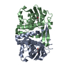
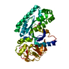
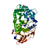
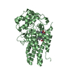
- PDB-5frv: crystal structure of the phenol-responsive sensory domain of the ... -
+ Open data

- Basic information
| Entry | Database: PDB / ID: 5frv | ||||||
|---|---|---|---|---|---|---|---|
| Title | crystal structure of the phenol-responsive sensory domain of the transcription activator PoxR in complex with 4-methylphenol (Cresol) | ||||||
 Components Components | Positive phenol-degradative gene regulator | ||||||
 Keywords Keywords | TRANSCRIPTION / PHENOL / POXR / DMPR / ENHANCER-BINDING PROTEIN / ACTIVATOR / ATPASE FAMILY | ||||||
| Function / homology |  Function and homology information Function and homology informationsequence-specific DNA binding / regulation of DNA-templated transcription / ATP binding / metal ion binding / identical protein binding Similarity search - Function | ||||||
| Biological species |  Ralstonia sp. E2 (bacteria) Ralstonia sp. E2 (bacteria) | ||||||
| Method |  X-RAY DIFFRACTION / SYNCHROTRON / MIR / Resolution: 1.9 Å X-RAY DIFFRACTION / SYNCHROTRON / MIR / Resolution: 1.9 Å | ||||||
 Authors Authors | Patil, V.V. / Woo, E.J. | ||||||
 Citation Citation | Journal: Structure / Year: 2016 Title: Structural Analysis of the Phenol-Responsive Sensory Domain of the Transcription Activator Poxr Authors: Patil, V.V. / Park, K.-H. / Lee, S.-G. / Woo, E.J. | ||||||
| History |
|
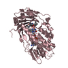
- Structure visualization
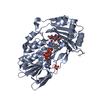
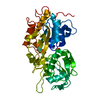
| Structure viewer | Molecule: MolmilJmol/JSmol |
|---|
- Downloads & links
-Download
| PDBx/mmCIF format | 5frv.cif.gz | 92.3 KB | Display | PDBx/mmCIF format |
|---|---|---|---|---|
| PDB format | pdb5frv.ent.gz | 70.4 KB | Display | PDB format |
| PDBx/mmJSON format | 5frv.json.gz | Tree view | PDBx/mmJSON format | |
| Others |  Other downloads Other downloads |
-Validation report
| Arichive directory | https://data.pdbj.org/pub/pdb/validation_reports/fr/5frvftp://data.pdbj.org/pub/pdb/validation_reports/fr/5frv | HTTPS FTP |
|---|
-Related structure data
| Related structure data |  5fruC  5frwC  5frxC  5fryC  5frzC  5fs0C C: citing same article ( |
|---|---|
| Similar structure data |


-Links
 PDBj
PDBj
- Assembly
| Deposited unit | 
| ||||||||
|---|---|---|---|---|---|---|---|---|---|
| 1 |
| ||||||||
| Unit cell |
|
-Components
| #1: Protein | Mass: 23495.096 Da / Num. of mol.: 2 / Fragment: PHENOL BINDING DOMAIN, UNP RESIDUES 1-211 Source method: isolated from a genetically manipulated source Source: (gene. exp.) Ralstonia sp. E2 (bacteria) / Gene: poxR / Variant: E2 / Plasmid: pET22B-CPD / Production host: #2: Chemical |   Mass: 65.409 Da / Num. of mol.: 2 / Source method: obtained synthetically / Formula: Zn Mass: 65.409 Da / Num. of mol.: 2 / Source method: obtained synthetically / Formula: Zn#3: Chemical | 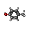  Mass: 108.138 Da / Num. of mol.: 2 / Source method: obtained synthetically / Formula: C7H8O Mass: 108.138 Da / Num. of mol.: 2 / Source method: obtained synthetically / Formula: C7H8O#4: Water | ChemComp-HOH / |  Mass: 18.015 Da / Num. of mol.: 122 / Source method: isolated from a natural source / Formula: H2O Mass: 18.015 Da / Num. of mol.: 122 / Source method: isolated from a natural source / Formula: H2O |
|---|
-Experimental details
-Experiment
| Experiment | Method: X-RAY DIFFRACTION |
|---|
- Sample preparation
| Crystal | Density Matthews: 1.84 Å3/Da / Density % sol: 33.22 % / Description: NONE |
|---|
-Data collection
| Diffraction | Mean temperature: 287 K |
|---|---|
| Diffraction source | Source: SYNCHROTRON / Site: PAL/PLS  / Beamline: 7A (6B, 6C1) / Wavelength: 0.97934 / Beamline: 7A (6B, 6C1) / Wavelength: 0.97934 |
| Detector | Type: ADSC CCD / Detector: CCD |
| Radiation | Protocol: SINGLE WAVELENGTH / Monochromatic (M) / Laue (L): M / Scattering type: x-ray |
| Radiation wavelength | Wavelength: 0.97934 Å / Relative weight: 1 |
| Reflection | Resolution: 1.5→50 Å / Num. obs: 56079 / % possible obs: 99.4 % / Observed criterion σ(I): -3 / Redundancy: 13.7 % / Rmerge(I) obs: 0.08 / Net I/σ(I): 37.29 |
| Reflection shell | Resolution: 1.5→1.53 Å / Redundancy: 12.3 % / Mean I/σ(I) obs: 0.43 / % possible all: 99.7 |
- Processing
| Software | Name: PHENIX / Version: (PHENIX.REFINE: 1.9_1692) / Classification: refinement | ||||||||||||||||||||||||||||||||||||||||||||||||||||||||
|---|---|---|---|---|---|---|---|---|---|---|---|---|---|---|---|---|---|---|---|---|---|---|---|---|---|---|---|---|---|---|---|---|---|---|---|---|---|---|---|---|---|---|---|---|---|---|---|---|---|---|---|---|---|---|---|---|---|
| Refinement | Method to determine structure: MIR Starting model: NONE Resolution: 1.9→23.348 Å / SU ML: 0.22 / σ(F): 1.33 / Phase error: 32.69 / Stereochemistry target values: ML
| ||||||||||||||||||||||||||||||||||||||||||||||||||||||||
| Solvent computation | Shrinkage radii: 0.9 Å / VDW probe radii: 1.11 Å / Solvent model: FLAT BULK SOLVENT MODEL | ||||||||||||||||||||||||||||||||||||||||||||||||||||||||
| Refinement step | Cycle: LAST / Resolution: 1.9→23.348 Å
| ||||||||||||||||||||||||||||||||||||||||||||||||||||||||
| Refine LS restraints |
| ||||||||||||||||||||||||||||||||||||||||||||||||||||||||
| LS refinement shell |
|
