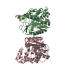
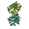
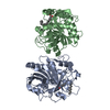
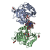
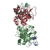
- PDB-5fqu: Orthorhombic crystal structure of of PlpD (selenomethionine deriv... -
+
Open data
ID or keywords:
Loading...
-
Basic information
Entry
Database: PDB / ID: 5fqu
Title
Orthorhombic crystal structure of of PlpD (selenomethionine derivative)
Components
PATATIN-LIKE PROTEIN, PLPD
Keywords
TRANSPORT PROTEIN / BACTERIAL SECRETION / PHOSPHOLIPASE / LIPID AFFINITY / INFECTION
Function / homology
Function and homology information
protein secretion by the type V secretion system / lipase activity / lipid catabolic process / cell outer membrane / extracellular region Similarity search - Function
Protocol: SINGLE WAVELENGTH / Monochromatic (M) / Laue (L): M / Scattering type: x-ray
Radiation wavelength
Wavelength: 0.97939 Å / Relative weight: 1
Reflection
Resolution: 2.74→85.58 Å / Num. obs: 19015 / % possible obs: 99.5 % / Observed criterion σ(I): 2.8 / Redundancy: 4.5 % / Biso Wilson estimate: 46.511 Å2 / Rsym value: 0.01 / Net I/σ(I): 10.42
Reflection shell
Resolution: 2.74→2.9 Å / Redundancy: 4.5 % / Mean I/σ(I) obs: 2.81 / Rsym value: 0.54 / % possible all: 96.5
-
Processing
Software
Name
Version
Classification
REFMAC
5.8.0135
refinement
XDS
datareduction
XSCALE
datascaling
autoSHARP
phasing
Refinement
Method to determine structure: SAD Starting model: NONE Resolution: 2.74→85.57 Å / Cor.coef. Fo:Fc: 0.965 / Cor.coef. Fo:Fc free: 0.943 / SU B: 19.334 / SU ML: 0.331 / Cross valid method: THROUGHOUT / ESU R: 0.629 / ESU R Free: 0.305 / Stereochemistry target values: MAXIMUM LIKELIHOOD Details: HYDROGENS HAVE BEEN ADDED IN THE RIDING POSITIONS. U VALUES REFINED INDIVIDUALLY
Rfactor
Num. reflection
% reflection
Selection details
Rfree
0.23801
968
5.1 %
RANDOM
Rwork
0.19579
-
-
-
obs
0.19805
17914
99.31 %
-
Solvent computation
Ion probe radii: 0.8 Å / Shrinkage radii: 0.8 Å / VDW probe radii: 1.2 Å / Solvent model: MASK
 Movie
Movie Controller

 Yorodumi
 Open data
 Basic information
 Components
 Keywords
 Function and homology information
 PSEUDOMONAS AERUGINOSA PAO1 (bacteria)
 X-RAY DIFFRACTION /
  Authors
 Citation
 Structure visualization
 Downloads & links
 Other downloads
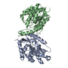


 PDBj
PDBj
 Assembly
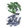

 Mass: 42.020 Da / Num. of mol.: 1 / Source method: obtained synthetically / Formula: N3
 Mass: 22.990 Da / Num. of mol.: 2 / Source method: obtained synthetically / Formula: Na
 Mass: 18.015 Da / Num. of mol.: 61 / Source method: isolated from a natural source / Formula: H2O
 Sample preparation
 / Beamline: ID29 / Wavelength: 0.97939
  Processing