 Movie
Movie Controller

[English] 日本語
 Yorodumi
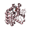
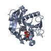
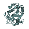
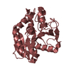
- PDB-1gt6: S146A mutant of Thermomyces (Humicola) lanuginosa lipase complex ... -
+ Open data

- Basic information
| Entry | Database: PDB / ID: 1gt6 | ||||||
|---|---|---|---|---|---|---|---|
| Title | S146A mutant of Thermomyces (Humicola) lanuginosa lipase complex with oleic acid | ||||||
 Components Components | Lipase | ||||||
 Keywords Keywords | LIPASE / HYDROLASE / LIPID DEGRADATION / ZYMOGEN | ||||||
| Function / homology |  Function and homology information Function and homology informationtriacylglycerol lipase / triacylglycerol lipase activity / lipid catabolic process Similarity search - Function | ||||||
| Biological species |   Thermomyces lanuginosus (fungus) Thermomyces lanuginosus (fungus) | ||||||
| Method |  X-RAY DIFFRACTION / SYNCHROTRON / MOLECULAR REPLACEMENT / Resolution: 2.2 Å X-RAY DIFFRACTION / SYNCHROTRON / MOLECULAR REPLACEMENT / Resolution: 2.2 Å | ||||||
 Authors Authors | Brzozowski, A.M. / Yapoudjian, S. / Ivanova, M.G. / Patkar, S.A. / Vind, J. / Svendsen, A. / Verger, R. | ||||||
 Citation Citation | Journal: Eur.J.Biochem. / Year: 2002 Title: Binding of Thermomyces (Humicola) lanuginosa lipase to the mixed micelles of cis-parinaric acid/NaTDC. Authors: Yapoudjian, S. / Ivanova, M.G. / Brzozowski, A.M. / Patkar, S.A. / Vind, J. / Svendsen, A. / Verger, R. | ||||||
| History |
|
- Structure visualization
| Structure viewer | Molecule: MolmilJmol/JSmol |
|---|
- Downloads & links
-Download
| PDBx/mmCIF format | 1gt6.cif.gz | 115.4 KB | Display | PDBx/mmCIF format |
|---|---|---|---|---|
| PDB format | pdb1gt6.ent.gz | 89.9 KB | Display | PDB format |
| PDBx/mmJSON format | 1gt6.json.gz | Tree view | PDBx/mmJSON format | |
| Others |  Other downloads Other downloads |
-Validation report
| Arichive directory | https://data.pdbj.org/pub/pdb/validation_reports/gt/1gt6ftp://data.pdbj.org/pub/pdb/validation_reports/gt/1gt6 | HTTPS FTP |
|---|
-Related structure data
| Related structure data |  1dt5S S: Starting model for refinement |
|---|---|
| Similar structure data |


-Links
 PDBj
PDBj


- Assembly
| Deposited unit | 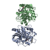
| ||||||||
|---|---|---|---|---|---|---|---|---|---|
| 1 | 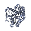
| ||||||||
| 2 | 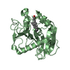
| ||||||||
| Unit cell |
|
-Components
| #1: Protein | Mass: 29326.484 Da / Num. of mol.: 2 / Mutation: S168A Source method: isolated from a genetically manipulated source Details: OLEIC ACID / Source: (gene. exp.) Thermomyces lanuginosus (fungus) / Gene: LIP / Production host: #2: Chemical |   Mass: 282.461 Da / Num. of mol.: 2 / Source method: obtained synthetically / Formula: C18H34O2 Mass: 282.461 Da / Num. of mol.: 2 / Source method: obtained synthetically / Formula: C18H34O2#3: Water | ChemComp-HOH / |  Mass: 18.015 Da / Num. of mol.: 169 / Source method: isolated from a natural source / Formula: H2O Mass: 18.015 Da / Num. of mol.: 169 / Source method: isolated from a natural source / Formula: H2OCompound details | ENGINEERED | Has protein modification | Y | |
|---|
-Experimental details
-Experiment
| Experiment | Method: X-RAY DIFFRACTION / Number of used crystals: 1 |
|---|
- Sample preparation
| Crystal | Density Matthews: 2.64 Å3/Da / Density % sol: 53 % | |||||||||||||||||||||||||||||||||||
|---|---|---|---|---|---|---|---|---|---|---|---|---|---|---|---|---|---|---|---|---|---|---|---|---|---|---|---|---|---|---|---|---|---|---|---|---|
| Crystal grow | pH: 8.51 Details: PH 8.5 TRIS BUFFER, 25 MM MGCL2, 25% W/V PEG 5K MME, 10 MM PROTEIN CONC 10-20 MG/ML IN 10 MM TRIS BUFFER PH 8.0 | |||||||||||||||||||||||||||||||||||
| Crystal grow | *PLUS pH: 8 / Method: unknown | |||||||||||||||||||||||||||||||||||
| Components of the solutions | *PLUS
|
-Data collection
| Diffraction | Mean temperature: 100 K |
|---|---|
| Diffraction source | Source: SYNCHROTRON / Site: ESRF  / Type: ESRF / Wavelength: 0.91 / Wavelength: 0.91 Å / Type: ESRF / Wavelength: 0.91 / Wavelength: 0.91 Å |
| Detector | Type: MARRESEARCH / Detector: CCD |
| Radiation | Protocol: SINGLE WAVELENGTH / Monochromatic (M) / Laue (L): M / Scattering type: x-ray |
| Radiation wavelength | Wavelength: 0.91 Å / Relative weight: 1 |
| Reflection | Resolution: 2.2→20 Å / Num. obs: 33614 / % possible obs: 97.8 % / Redundancy: 3.5 % / Rsym value: 0.075 / Net I/σ(I): 9.3 |
| Reflection shell | Resolution: 2.2→2.28 Å / Redundancy: 3 % / Mean I/σ(I) obs: 2.5 / Rsym value: 0.5 / % possible all: 94.7 |
| Reflection | *PLUS Highest resolution: 2.2 Å / Rmerge(I) obs: 0.075 |
| Reflection shell | *PLUS Highest resolution: 2.2 Å / % possible obs: 96.2 % / Rmerge(I) obs: 0.44 / Mean I/σ(I) obs: 3.2 |
- Processing
| Software |
| ||||||||||||||||||||
|---|---|---|---|---|---|---|---|---|---|---|---|---|---|---|---|---|---|---|---|---|---|
| Refinement | Method to determine structure: MOLECULAR REPLACEMENT Starting model: PDB ENTRY 1DT5 Resolution: 2.2→20 Å / SU B: 5.96346 / SU ML: 0.15322 / Cross valid method: THROUGHOUT / σ(F): 0 / ESU R: 0.27451 / ESU R Free: 0.20358 Details: OLEIC ACID IN MOLECULE B IS COMPLETE B IN MOLECULE A ALKYL CHAIN IS VISIBLE TILL C10 CARBON SO THE OCCUPANCY OF THE C11-C18 WERE SET TO 0 (BUT MODELLED).
| ||||||||||||||||||||
| Displacement parameters | Biso mean: 21.158 Å2
| ||||||||||||||||||||
| Refinement step | Cycle: LAST / Resolution: 2.2→20 Å
| ||||||||||||||||||||
| Refinement | *PLUS Highest resolution: 2.2 Å / Lowest resolution: 20 Å / Rfactor Rfree: 0.239 / Rfactor Rwork: 0.214 | ||||||||||||||||||||
| Solvent computation | *PLUS | ||||||||||||||||||||
| Displacement parameters | *PLUS | ||||||||||||||||||||
| Refine LS restraints | *PLUS
|
