 Movie
Movie Controller

[English] 日本語
 Yorodumi
- PDB-1dt3: THE STRUCTURAL ORIGINS OF INTERFACIAL ACTIVATION IN THERMOMYCES (... -
+ Open data

- Basic information
| Entry | Database: PDB / ID: 1dt3 | ||||||
|---|---|---|---|---|---|---|---|
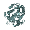
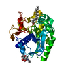
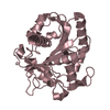
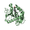
| Title | THE STRUCTURAL ORIGINS OF INTERFACIAL ACTIVATION IN THERMOMYCES (HUMICOLA) LANUGINOSA LIPASE | ||||||
 Components Components | LIPASE | ||||||
 Keywords Keywords | HYDROLASE / lipase / thermomyces linuginosa / interfacial activation | ||||||
| Function / homology |  Function and homology information Function and homology informationtriacylglycerol lipase / triacylglycerol lipase activity / lipid catabolic process Similarity search - Function | ||||||
| Biological species |   Thermomyces lanuginosus (fungus) Thermomyces lanuginosus (fungus) | ||||||
| Method |  X-RAY DIFFRACTION / Resolution: 2.6 Å X-RAY DIFFRACTION / Resolution: 2.6 Å | ||||||
 Authors Authors | Brozozowski, A.M. / Savage, H. | ||||||
 Citation Citation | Journal: Biochemistry / Year: 2000 Title: Structural origins of the interfacial activation in Thermomyces (Humicola) lanuginosa lipase. Authors: Brzozowski, A.M. / Savage, H. / Verma, C.S. / Turkenburg, J.P. / Lawson, D.M. / Svendsen, A. / Patkar, S. | ||||||
| History |
|
- Structure visualization
| Structure viewer | Molecule: MolmilJmol/JSmol |
|---|
- Downloads & links
-Download
| PDBx/mmCIF format | 1dt3.cif.gz | 117.7 KB | Display | PDBx/mmCIF format |
|---|---|---|---|---|
| PDB format | pdb1dt3.ent.gz | 92.2 KB | Display | PDB format |
| PDBx/mmJSON format | 1dt3.json.gz | Tree view | PDBx/mmJSON format | |
| Others |  Other downloads Other downloads |
-Validation report
| Arichive directory | https://data.pdbj.org/pub/pdb/validation_reports/dt/1dt3ftp://data.pdbj.org/pub/pdb/validation_reports/dt/1dt3 | HTTPS FTP |
|---|
-Related structure data


-Links
 PDBj
PDBj- Assembly
| Deposited unit | 
| ||||||||
|---|---|---|---|---|---|---|---|---|---|
| 1 | 
| ||||||||
| 2 | 
| ||||||||
| Unit cell |
| ||||||||
| Details | The biological assembly is a monomer |
-Components
| #1: Protein | Mass: 29342.484 Da / Num. of mol.: 2 / Source method: isolated from a natural source / Source: (natural) Thermomyces lanuginosus (fungus) / References: UniProt: O59952, triacylglycerol lipase#2: Water | ChemComp-HOH / |  Mass: 18.015 Da / Num. of mol.: 242 / Source method: isolated from a natural source / Formula: H2O Mass: 18.015 Da / Num. of mol.: 242 / Source method: isolated from a natural source / Formula: H2OHas protein modification | Y | |
|---|
-Experimental details
-Experiment
| Experiment | Method: X-RAY DIFFRACTION / Number of used crystals: 1 |
|---|
- Sample preparation
| Crystal | Density Matthews: 3.88 Å3/Da / Density % sol: 68.33 % | |||||||||||||||||||||||||
|---|---|---|---|---|---|---|---|---|---|---|---|---|---|---|---|---|---|---|---|---|---|---|---|---|---|---|
| Crystal grow | Temperature: 279 K / Method: vapor diffusion, hanging drop / pH: 8.1 Details: PEG 5000, magnesium chloride, , pH 8.1, VAPOR DIFFUSION, HANGING DROP, temperature 6K | |||||||||||||||||||||||||
| Crystal grow | *PLUS Method: unknown | |||||||||||||||||||||||||
| Components of the solutions | *PLUS
|
-Data collection
| Diffraction | Mean temperature: 110 K |
|---|---|
| Diffraction source | Source: ROTATING ANODE / Type: RIGAKU RU200 / Wavelength: 1.5418 |
| Detector | Type: MARRESEARCH / Detector: IMAGE PLATE / Date: Mar 6, 1999 |
| Radiation | Protocol: SINGLE WAVELENGTH / Monochromatic (M) / Laue (L): M / Scattering type: x-ray |
| Radiation wavelength | Wavelength: 1.5418 Å / Relative weight: 1 |
| Reflection | Resolution: 2.6→20 Å / Num. all: 27814 / Num. obs: 26160 / % possible obs: 94 % / Observed criterion σ(F): 2 / Observed criterion σ(I): 2 / Redundancy: 5 % / Biso Wilson estimate: 55 Å2 / Rmerge(I) obs: 0.052 / Net I/σ(I): 26 |
| Reflection shell | Resolution: 2.6→20 Å / Redundancy: 26 % / Rmerge(I) obs: 0.048 / Num. unique all: 26160 / % possible all: 99.6 |
| Reflection | *PLUS Highest resolution: 2.5 Å / Lowest resolution: 24.5 Å / Num. obs: 31111 / % possible obs: 98.8 % / Redundancy: 4.7 % |
| Reflection shell | *PLUS Highest resolution: 2.5 Å / Lowest resolution: 2.54 Å / % possible obs: 95.3 % / Redundancy: 4 % / Rmerge(I) obs: 0.155 / Mean I/σ(I) obs: 9.4 |
- Processing
| Software |
| |||||||||||||||||||||||||
|---|---|---|---|---|---|---|---|---|---|---|---|---|---|---|---|---|---|---|---|---|---|---|---|---|---|---|
| Refinement | Resolution: 2.6→20 Å / σ(F): 0 / σ(I): 0 Stereochemistry target values: rms bond = 0.02A rms angle = 0.06A Details: rms bond = 0.013A rms angle = 0.041A
| |||||||||||||||||||||||||
| Refinement step | Cycle: LAST / Resolution: 2.6→20 Å
| |||||||||||||||||||||||||
| Refine LS restraints |
| |||||||||||||||||||||||||
| Software | *PLUS Name: REFMAC / Classification: refinement | |||||||||||||||||||||||||
| Refinement | *PLUS Rfactor Rfree: 0.372 / Rfactor Rwork: 0.296 | |||||||||||||||||||||||||
| Solvent computation | *PLUS | |||||||||||||||||||||||||
| Displacement parameters | *PLUS Biso mean: 61.2 Å2 | |||||||||||||||||||||||||
| Refine LS restraints | *PLUS
|
