- PDB-5fen: Crystal structure of the Q108K:K40L:T53C mutant of human Cellular... -
+
Open data
ID or keywords:
Loading...
-
Basic information
Entry
Database: PDB / ID: 5fen
Title
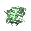
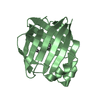
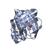
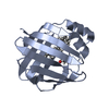
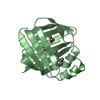
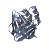
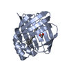
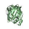
Crystal structure of the Q108K:K40L:T53C mutant of human Cellular Retinol Binding Protein II in complex with All-trans-Retinal after 24 hours of incubation at 1.55 Angstrom Resolution
Components
Retinol-binding protein 2
Keywords
TRANSPORT PROTEIN / all-trans retinal / retinal light absorbing pigment / wavelength regulation
Function / homology
Function and homology information
vitamin A metabolic process / triglyceride biosynthetic process / retinoid binding / retinal binding / molecular carrier activity / retinol binding / epidermis development / fatty acid transport / Retinoid metabolism and transport / fatty acid binding ...vitamin A metabolic process / triglyceride biosynthetic process / retinoid binding / retinal binding / molecular carrier activity / retinol binding / epidermis development / fatty acid transport / Retinoid metabolism and transport / fatty acid binding / nucleus / cytosol Similarity search - Function
In the structure databanks used in Yorodumi, some data are registered as the other names, "COVID-19 virus" and "2019-nCoV". Here are the details of the virus and the list of structure data.
Jan 31, 2019. EMDB accession codes are about to change! (news from PDBe EMDB page)
EMDB accession codes are about to change! (news from PDBe EMDB page)
The allocation of 4 digits for EMDB accession codes will soon come to an end. Whilst these codes will remain in use, new EMDB accession codes will include an additional digit and will expand incrementally as the available range of codes is exhausted. The current 4-digit format prefixed with “EMD-” (i.e. EMD-XXXX) will advance to a 5-digit format (i.e. EMD-XXXXX), and so on. It is currently estimated that the 4-digit codes will be depleted around Spring 2019, at which point the 5-digit format will come into force.
The EM Navigator/Yorodumi systems omit the EMD- prefix.
Related info.:Q: What is EMD? / ID/Accession-code notation in Yorodumi/EM Navigator
Yorodumi is a browser for structure data from EMDB, PDB, SASBDB, etc.
This page is also the successor to EM Navigator detail page, and also detail information page/front-end page for Omokage search.
The word "yorodu" (or yorozu) is an old Japanese word meaning "ten thousand". "mi" (miru) is to see.
Related info.:EMDB / PDB / SASBDB / Comparison of 3 databanks / Yorodumi Search / Aug 31, 2016. New EM Navigator & Yorodumi / Yorodumi Papers / Jmol/JSmol / Function and homology information / Changes in new EM Navigator and Yorodumi
 Movie
Movie Controller

 Yorodumi
 Open data
 Basic information
 Components
 Keywords
 Function and homology information
 Homo sapiens (human)
 X-RAY DIFFRACTION /
  Authors
 Citation
 Structure visualization
 Downloads & links
 Other downloads


 PDBj
PDBj


 Assembly


 Mass: 284.436 Da / Num. of mol.: 2 / Source method: obtained synthetically / Formula: C20H28O
 Mass: 59.044 Da / Num. of mol.: 2 / Source method: obtained synthetically / Formula: C2H3O2
 Mass: 18.015 Da / Num. of mol.: 209 / Source method: isolated from a natural source / Formula: H2O
 Sample preparation
 / Beamline: 21-ID-D / Wavelength: 0.9786 Å
 Processing