[English] 日本語
Yorodumi
- PDB-5fc9: Novel Purple Cupredoxin from Nitrosopumilus maritimus -

+
Open data


ID or keywords:

Loading...

-
Basic information

Entry
Database: PDB / ID: 5fc9
TitleNovel Purple Cupredoxin from Nitrosopumilus maritimus
ComponentsBlue (Type 1) copper domain protein
KeywordsMETAL BINDING PROTEIN / Cupredoxin / NO oxidation / nitrogen cycle / open type 1 copper site
Function / homology
Function and homology information


electron transfer activity / copper ion binding
Similarity search - Function
Blue (type 1) copper domain / Copper binding proteins, plastocyanin/azurin family / Cupredoxins - blue copper proteins / Cupredoxin / Immunoglobulin-like / Sandwich / Mainly Beta
Similarity search - Domain/homology
COPPER (II) ION / Blue (Type 1) copper domain protein
Similarity search - Component
Biological speciesNitrosopumilus maritimus (archaea)
MethodX-RAY DIFFRACTION / SYNCHROTRON / MOLECULAR REPLACEMENT / Resolution: 1.6 Å
AuthorsHosseinzadeh, P. / Lu, Y. / Robinson, H. / Gao, Y.-G.
Funding support United States, 1items
OrganizationGrant numberCountry
National Science Foundation (NSF, United States)CHE-1413328 United States
CitationJournal: J.Am.Chem.Soc. / Year: 2016
Title: A Purple Cupredoxin from Nitrosopumilus maritimus Containing a Mononuclear Type 1 Copper Center with an Open Binding Site.
Authors: Hosseinzadeh, P. / Tian, S. / Marshall, N.M. / Hemp, J. / Mullen, T. / Nilges, M.J. / Gao, Y.G. / Robinson, H. / Stahl, D.A. / Gennis, R.B. / Lu, Y.
History
DepositionDec 15, 2015Deposition site: RCSB / Processing site: RCSB
Revision 1.0May 11, 2016Provider: repository / Type: Initial release
Revision 1.1Jun 8, 2016Group: Database references
Revision 1.2Sep 20, 2017Group: Author supporting evidence / Derived calculations / Category: pdbx_audit_support / pdbx_struct_oper_list
Item: _pdbx_audit_support.funding_organization / _pdbx_struct_oper_list.symmetry_operation
Revision 1.3Nov 27, 2019Group: Author supporting evidence / Category: pdbx_audit_support / Item: _pdbx_audit_support.funding_organization
Revision 1.4Sep 27, 2023Group: Data collection / Database references ...Data collection / Database references / Derived calculations / Refinement description
Category: chem_comp_atom / chem_comp_bond ...chem_comp_atom / chem_comp_bond / database_2 / pdbx_initial_refinement_model / struct_conn
Item: _database_2.pdbx_DOI / _database_2.pdbx_database_accession ..._database_2.pdbx_DOI / _database_2.pdbx_database_accession / _struct_conn.pdbx_dist_value / _struct_conn.ptnr1_auth_asym_id / _struct_conn.ptnr1_auth_comp_id / _struct_conn.ptnr1_auth_seq_id / _struct_conn.ptnr1_label_asym_id / _struct_conn.ptnr1_label_atom_id / _struct_conn.ptnr1_label_comp_id / _struct_conn.ptnr1_label_seq_id / _struct_conn.ptnr1_symmetry / _struct_conn.ptnr2_auth_asym_id / _struct_conn.ptnr2_auth_comp_id / _struct_conn.ptnr2_auth_seq_id / _struct_conn.ptnr2_label_asym_id / _struct_conn.ptnr2_label_atom_id / _struct_conn.ptnr2_label_comp_id / _struct_conn.ptnr2_label_seq_id / _struct_conn.ptnr2_symmetry

-
Structure visualization

Structure viewerMolecule:
MolmilJmol/JSmol

Downloads & links

-
Assembly

Deposited unit
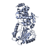
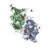
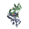
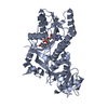
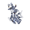
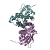
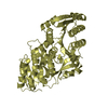
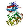
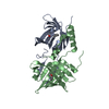
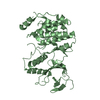
A: Blue (Type 1) copper domain protein
B: Blue (Type 1) copper domain protein
C: Blue (Type 1) copper domain protein
D: Blue (Type 1) copper domain protein
hetero molecules


Theoretical massNumber of molelcules
Total (without water)42,8099
Polymers42,4914
Non-polymers3185
Water10,485582
1


  • Idetical with deposited unit
  • defined by author&software
TypeNameSymmetry operationNumber
identity operation1_555x,y,z1
Buried area4920 Å2
ΔGint-92 kcal/mol
Surface area15910 Å2
MethodPISA
Unit cell
Length a, b, c (Å)68.887, 68.887, 184.450
Angle α, β, γ (deg.)90.00, 90.00, 90.00
Int Tables number92
Space group name H-MP41212

-
Components

#1: Protein
Blue (Type 1) copper domain protein


Mass: 10622.711 Da / Num. of mol.: 4
Source method: isolated from a genetically manipulated source
Details: Protein containing Cu ions
Source: (gene. exp.) Nitrosopumilus maritimus (strain SCM1) (archaea)
Strain: SCM1 / Gene: Nmar_1307 / Plasmid: pET22b / Details (production host): periplasmic expression / Production host: Escherichia coli (E. coli) / Strain (production host): Rosetta / References: UniProt: A9A2G4
#2: Chemical
ChemComp-CU / COPPER (II) ION / Copper


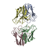
Mass: 63.546 Da / Num. of mol.: 5 / Source method: obtained synthetically / Formula: Cu
#3: Water ChemComp-HOH / water / Water


Mass: 18.015 Da / Num. of mol.: 582 / Source method: isolated from a natural source / Formula: H2O

-
Experimental details

-
Experiment

ExperimentMethod: X-RAY DIFFRACTION

-
Sample preparation

CrystalDensity Matthews: 2.58 Å3/Da / Density % sol: 52.31 %
Crystal growTemperature: 277 K / Method: vapor diffusion, hanging drop / pH: 8
Details: Crystals of N. mar_1307 were grown by the hanging drop vapor diffusion method. Protein solution was prepared in 50 mM sodium acetate buffer at pH 6.0 to a concentration of about 2 mM. A 2 uL ...Details: Crystals of N. mar_1307 were grown by the hanging drop vapor diffusion method. Protein solution was prepared in 50 mM sodium acetate buffer at pH 6.0 to a concentration of about 2 mM. A 2 uL portion of this protein solution was then mixed with 2 uL of a well buffer solution consisting of 0.1 M TrisHCl at pH 8.0, 20mM CuSO4, 0.1M LiNO3 and varying amounts of polyethylene glycol (PEG). The highest quality crystals formed from wells with 35 % w/v PEG 4000 after about 2 months at 4 C

-
Data collection

DiffractionMean temperature: 80 K
Diffraction sourceSource: SYNCHROTRON / Site: NSLS / Beamline: X29A / Wavelength: 1.075 Å
DetectorType: ADSC QUANTUM 315r / Detector: CCD / Date: Apr 27, 2011
RadiationProtocol: SINGLE WAVELENGTH / Monochromatic (M) / Laue (L): M / Scattering type: x-ray
Radiation wavelengthWavelength: 1.075 Å / Relative weight: 1
ReflectionResolution: 1.6→50 Å / Num. obs: 59707 / % possible obs: 100 % / Redundancy: 29.5 % / Rmerge(I) obs: 0.105 / Χ2: 1.038 / Net I/av σ(I): 36.56 / Net I/σ(I): 11 / Num. measured all: 1760680
Reflection shell

Diffraction-ID: 1 / Rejects: 0

Resolution (Å)Redundancy (%)Rmerge(I) obsNum. unique allΧ2% possible all
1.6-1.6628.10.71958370.879100
1.66-1.7228.30.55658580.916100
1.72-1.828.40.41158620.964100
1.8-1.928.50.29658891.044100
1.9-2.0228.60.20358971.083100
2.02-2.1728.50.14759391.069100
2.17-2.3928.50.11959471.012100
2.39-2.7428.30.10659921.08100
2.74-3.4527.70.08760801.127100
3.45-5039.20.08764061.14499.9

-
Processing

Software
NameVersionClassification
PHENIX1.9_1692refinement
HKL-2000data reduction
HKL-2000data scaling
PHENIXphasing
RefinementMethod to determine structure: MOLECULAR REPLACEMENT
Starting model: 4AZU
Resolution: 1.6→45.87 Å / SU ML: 0.16 / Cross valid method: FREE R-VALUE / σ(F): 1.34 / Phase error: 19.18 / Stereochemistry target values: ML
RfactorNum. reflection% reflection
Rfree0.1978 3747 3.35 %
Rwork0.1731 --
obs0.174 -99.96 %
Solvent computationShrinkage radii: 0.9 Å / VDW probe radii: 1.11 Å / Solvent model: FLAT BULK SOLVENT MODEL
Refinement stepCycle: LAST / Resolution: 1.6→45.87 Å
ProteinNucleic acidLigandSolventTotal
Num. atoms3012 0 5 582 3599
Refine LS restraints
Refine-IDTypeDev idealNumber
X-RAY DIFFRACTIONf_bond_d0.0063104
X-RAY DIFFRACTIONf_angle_d1.0684264
X-RAY DIFFRACTIONf_dihedral_angle_d12.6351080
X-RAY DIFFRACTIONf_chiral_restr0.043468
X-RAY DIFFRACTIONf_plane_restr0.006564
LS refinement shell
Resolution (Å)Rfactor RfreeNum. reflection RfreeRfactor RworkNum. reflection RworkRefine-ID% reflection obs (%)
1.5995-1.61980.24041400.20843934X-RAY DIFFRACTION99
1.6198-1.64110.28351390.20574027X-RAY DIFFRACTION100
1.6411-1.66360.20741370.19813958X-RAY DIFFRACTION100
1.6636-1.68730.22061410.19594048X-RAY DIFFRACTION100
1.6873-1.71250.24821360.20484004X-RAY DIFFRACTION100
1.7125-1.73930.24151450.2144022X-RAY DIFFRACTION100
1.7393-1.76780.25811430.20364041X-RAY DIFFRACTION100
1.7678-1.79830.23911400.20293952X-RAY DIFFRACTION100
1.7983-1.8310.26371420.19944016X-RAY DIFFRACTION100
1.831-1.86620.22541340.19974004X-RAY DIFFRACTION100
1.8662-1.90430.25051380.19564003X-RAY DIFFRACTION100
1.9043-1.94570.22671390.18024015X-RAY DIFFRACTION100
1.9457-1.9910.2021370.18374017X-RAY DIFFRACTION100
1.991-2.04080.20531380.18074002X-RAY DIFFRACTION100
2.0408-2.09590.19241390.17384012X-RAY DIFFRACTION100
2.0959-2.15760.17581380.17264017X-RAY DIFFRACTION100
2.1576-2.22720.18151370.1754013X-RAY DIFFRACTION100
2.2272-2.30680.18711380.16994007X-RAY DIFFRACTION100
2.3068-2.39920.23091430.17364000X-RAY DIFFRACTION100
2.3992-2.50840.20191380.18883993X-RAY DIFFRACTION100
2.5084-2.64060.22591370.19054054X-RAY DIFFRACTION100
2.6406-2.8060.21141360.18633960X-RAY DIFFRACTION100
2.806-3.02270.21161370.17584045X-RAY DIFFRACTION100
3.0227-3.32680.16631350.16993989X-RAY DIFFRACTION100
3.3268-3.8080.18091400.14894025X-RAY DIFFRACTION100
3.808-4.79680.14391400.14024005X-RAY DIFFRACTION100
4.7968-45.88920.19641400.16324005X-RAY DIFFRACTION100

+
About Yorodumi

-
News

-
Feb 9, 2022. New format data for meta-information of EMDB entries

New format data for meta-information of EMDB entries

  • Version 3 of the EMDB header file is now the official format.
  • The previous official version 1.9 will be removed from the archive.

Related info.:EMDB header

External links:wwPDB to switch to version 3 of the EMDB data model

-
Aug 12, 2020. Covid-19 info

Covid-19 info

URL: https://pdbj.org/emnavi/covid19.php

New page: Covid-19 featured information page in EM Navigator.

Related info.:Covid-19 info / Mar 5, 2020. Novel coronavirus structure data

+
Mar 5, 2020. Novel coronavirus structure data

Novel coronavirus structure data

Related info.:Yorodumi Speices / Aug 12, 2020. Covid-19 info

External links:COVID-19 featured content - PDBj / Molecule of the Month (242):Coronavirus Proteases

+
Jan 31, 2019. EMDB accession codes are about to change! (news from PDBe EMDB page)

EMDB accession codes are about to change! (news from PDBe EMDB page)

  • The allocation of 4 digits for EMDB accession codes will soon come to an end. Whilst these codes will remain in use, new EMDB accession codes will include an additional digit and will expand incrementally as the available range of codes is exhausted. The current 4-digit format prefixed with “EMD-” (i.e. EMD-XXXX) will advance to a 5-digit format (i.e. EMD-XXXXX), and so on. It is currently estimated that the 4-digit codes will be depleted around Spring 2019, at which point the 5-digit format will come into force.
  • The EM Navigator/Yorodumi systems omit the EMD- prefix.

Related info.:Q: What is EMD? / ID/Accession-code notation in Yorodumi/EM Navigator

External links:EMDB Accession Codes are Changing Soon! / Contact to PDBj

+
Jul 12, 2017. Major update of PDB

Major update of PDB

  • wwPDB released updated PDB data conforming to the new PDBx/mmCIF dictionary.
  • This is a major update changing the version number from 4 to 5, and with Remediation, in which all the entries are updated.
  • In this update, many items about electron microscopy experimental information are reorganized (e.g. em_software).
  • Now, EM Navigator and Yorodumi are based on the updated data.

External links:wwPDB Remediation / Enriched Model Files Conforming to OneDep Data Standards Now Available in the PDB FTP Archive

-
Yorodumi

Thousand views of thousand structures

  • Yorodumi is a browser for structure data from EMDB, PDB, SASBDB, etc.
  • This page is also the successor to EM Navigator detail page, and also detail information page/front-end page for Omokage search.
  • The word "yorodu" (or yorozu) is an old Japanese word meaning "ten thousand". "mi" (miru) is to see.

Related info.:EMDB / PDB / SASBDB / Comparison of 3 databanks / Yorodumi Search / Aug 31, 2016. New EM Navigator & Yorodumi / Yorodumi Papers / Jmol/JSmol / Function and homology information / Changes in new EM Navigator and Yorodumi

Read more