 Movie
Movie Controller

[English] 日本語
 Yorodumi
- PDB-5fa9: Bifunctional Methionine Sulfoxide Reductase AB (MsrAB) from Trepo... -
+ Open data

- Basic information
| Entry | Database: PDB / ID: 5fa9 | ||||||
|---|---|---|---|---|---|---|---|
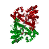
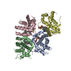
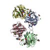
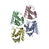
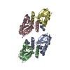
| Title | Bifunctional Methionine Sulfoxide Reductase AB (MsrAB) from Treponema denticola | ||||||
 Components Components | Peptide methionine sulfoxide reductase MsrA | ||||||
 Keywords Keywords | OXIDOREDUCTASE / Methionine sulfoxide reductase(Msr) / fusion protein / Protein oxidation | ||||||
| Function / homology |  Function and homology information Function and homology informationpeptide-methionine (R)-S-oxide reductase activity / L-methionine (S)-S-oxide reductase activity / peptide-methionine (S)-S-oxide reductase / peptide-methionine (S)-S-oxide reductase activity / protein repair / protein modification process / response to oxidative stress / cytoplasm Similarity search - Function | ||||||
| Biological species |  Treponema denticola ATCC 35405 (bacteria) Treponema denticola ATCC 35405 (bacteria) | ||||||
| Method |  X-RAY DIFFRACTION / SYNCHROTRON / MOLECULAR REPLACEMENT / Resolution: 2.302 Å X-RAY DIFFRACTION / SYNCHROTRON / MOLECULAR REPLACEMENT / Resolution: 2.302 Å | ||||||
 Authors Authors | Han, A. / Son, J. / Kim, H.-Y. / Hwang, K.Y. | ||||||
 Citation Citation | Journal: Biochemistry / Year: 2016 Title: Essential Role of the Linker Region in the Higher Catalytic Efficiency of a Bifunctional MsrA-MsrB Fusion Protein Authors: Han, A.R. / Kim, M.J. / Kwak, G.H. / Son, J. / Hwang, K.Y. / Kim, H.Y. | ||||||
| History |
|
- Structure visualization
| Structure viewer | Molecule: MolmilJmol/JSmol |
|---|
- Downloads & links
-Download
| PDBx/mmCIF format | 5fa9.cif.gz | 139.5 KB | Display | PDBx/mmCIF format |
|---|---|---|---|---|
| PDB format | pdb5fa9.ent.gz | 107.9 KB | Display | PDB format |
| PDBx/mmJSON format | 5fa9.json.gz | Tree view | PDBx/mmJSON format | |
| Others |  Other downloads Other downloads |
-Validation report
| Arichive directory | https://data.pdbj.org/pub/pdb/validation_reports/fa/5fa9ftp://data.pdbj.org/pub/pdb/validation_reports/fa/5fa9 | HTTPS FTP |
|---|
-Related structure data
| Related structure data | 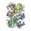 3e0mS S: Starting model for refinement |
|---|---|
| Similar structure data |


-Links
 PDBj
PDBj


- Assembly
| Deposited unit | 
| ||||||||
|---|---|---|---|---|---|---|---|---|---|
| 1 |
| ||||||||
| Unit cell |
|
-Components
| #1: Protein | Mass: 36907.484 Da / Num. of mol.: 2 / Mutation: C11S, C285S Source method: isolated from a genetically manipulated source Source: (gene. exp.) Treponema denticola ATCC 35405 (bacteria)Strain: ATCC 35405 / Gene: msrA, TDE_0709 / Plasmid: pET21b / Production host: References: UniProt: Q73PT7, peptide-methionine (S)-S-oxide reductase #2: Chemical |   Mass: 152.235 Da / Num. of mol.: 2 / Source method: obtained synthetically / Formula: C4H8O2S2 Mass: 152.235 Da / Num. of mol.: 2 / Source method: obtained synthetically / Formula: C4H8O2S2#3: Chemical | ChemComp-DTT /   Mass: 154.251 Da / Num. of mol.: 5 / Source method: obtained synthetically / Formula: C4H10O2S2 Mass: 154.251 Da / Num. of mol.: 5 / Source method: obtained synthetically / Formula: C4H10O2S2#4: Water | ChemComp-HOH / |  Mass: 18.015 Da / Num. of mol.: 241 / Source method: isolated from a natural source / Formula: H2O Mass: 18.015 Da / Num. of mol.: 241 / Source method: isolated from a natural source / Formula: H2O |
|---|
-Experimental details
-Experiment
| Experiment | Method: X-RAY DIFFRACTION / Number of used crystals: 1 |
|---|
- Sample preparation
| Crystal | Density Matthews: 3.43 Å3/Da / Density % sol: 64.1 % |
|---|---|
| Crystal grow | Temperature: 293 K / Method: vapor diffusion, sitting drop / pH: 6.2 Details: 0.2 M sodium chloride, 0.1 M sodium potassium phosphate buffer (pH 6.2), 50% polyethylene glycol 200 PH range: 6.0-6.4 |
-Data collection
| Diffraction | Mean temperature: 100 K |
|---|---|
| Diffraction source | Source: SYNCHROTRON / Site: PAL/PLS  / Beamline: 5C (4A) / Wavelength: 0.9796 Å / Beamline: 5C (4A) / Wavelength: 0.9796 Å |
| Detector | Type: ADSC QUANTUM 315r / Detector: CCD / Date: Apr 11, 2015 |
| Radiation | Protocol: SINGLE WAVELENGTH / Monochromatic (M) / Laue (L): M / Scattering type: x-ray |
| Radiation wavelength | Wavelength: 0.9796 Å / Relative weight: 1 |
| Reflection | Resolution: 2.3→30 Å / Num. obs: 42971 / % possible obs: 97.26 % / Redundancy: 4.6 % / Rmerge(I) obs: 0.081 / Net I/σ(I): 23.2 |
| Reflection shell | Resolution: 2.3→2.34 Å / Redundancy: 2.9 % / Rmerge(I) obs: 0.353 / Mean I/σ(I) obs: 2.37 / % possible all: 92.7 |
- Processing
| Software |
| |||||||||||||||||||||||||||||||||||||||||||||||||||||||||||||||||||||||||||||||||||||||||||||||||||||||||
|---|---|---|---|---|---|---|---|---|---|---|---|---|---|---|---|---|---|---|---|---|---|---|---|---|---|---|---|---|---|---|---|---|---|---|---|---|---|---|---|---|---|---|---|---|---|---|---|---|---|---|---|---|---|---|---|---|---|---|---|---|---|---|---|---|---|---|---|---|---|---|---|---|---|---|---|---|---|---|---|---|---|---|---|---|---|---|---|---|---|---|---|---|---|---|---|---|---|---|---|---|---|---|---|---|---|---|
| Refinement | Method to determine structure: MOLECULAR REPLACEMENT Starting model: 3E0M Resolution: 2.302→29.634 Å / SU ML: 0.31 / Cross valid method: FREE R-VALUE / σ(F): 1.51 / Phase error: 23.92 / Stereochemistry target values: ML
| |||||||||||||||||||||||||||||||||||||||||||||||||||||||||||||||||||||||||||||||||||||||||||||||||||||||||
| Solvent computation | Shrinkage radii: 0.9 Å / VDW probe radii: 1.11 Å / Solvent model: FLAT BULK SOLVENT MODEL | |||||||||||||||||||||||||||||||||||||||||||||||||||||||||||||||||||||||||||||||||||||||||||||||||||||||||
| Refinement step | Cycle: LAST / Resolution: 2.302→29.634 Å
| |||||||||||||||||||||||||||||||||||||||||||||||||||||||||||||||||||||||||||||||||||||||||||||||||||||||||
| Refine LS restraints |
| |||||||||||||||||||||||||||||||||||||||||||||||||||||||||||||||||||||||||||||||||||||||||||||||||||||||||
| LS refinement shell |
|
