 Movie
Movie Controller

[English] 日本語
 Yorodumi
- PDB-5f05: Crystal structure of glutathione transferase F5 from Populus tric... -
+ Open data

- Basic information
| Entry | Database: PDB / ID: 5f05 | ||||||
|---|---|---|---|---|---|---|---|
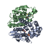
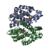
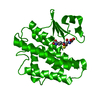
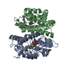
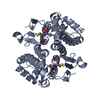
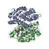
| Title | Crystal structure of glutathione transferase F5 from Populus trichocarpa | ||||||
 Components Components | Phi class glutathione transferase GSTF5 | ||||||
 Keywords Keywords | TRANSFERASE / glutathione / ligandin | ||||||
| Function / homology |  Function and homology information Function and homology informationtoxin catabolic process / glutathione binding / glutathione transferase / glutathione transferase activity / glutathione metabolic process / metal ion binding / cytosol / cytoplasm Similarity search - Function | ||||||
| Biological species |  | ||||||
| Method |  X-RAY DIFFRACTION / SYNCHROTRON / MOLECULAR REPLACEMENT / Resolution: 1.7 Å X-RAY DIFFRACTION / SYNCHROTRON / MOLECULAR REPLACEMENT / Resolution: 1.7 Å | ||||||
 Authors Authors | Didierjean, C. / Rouhier, N. / Pegeot, H. / Gense, F. | ||||||
| Funding support |  France, 1items France, 1items
| ||||||
 Citation Citation | Journal: FEBS J. / Year: 2017 Title: Structural plasticity among glutathione transferase Phi members: natural combination of catalytic residues confers dual biochemical activities. Authors: Pegeot, H. / Mathiot, S. / Perrot, T. / Gense, F. / Hecker, A. / Didierjean, C. / Rouhier, N. | ||||||
| History |
|
- Structure visualization
| Structure viewer | Molecule: MolmilJmol/JSmol |
|---|
- Downloads & links
-Download
| PDBx/mmCIF format | 5f05.cif.gz | 344.9 KB | Display | PDBx/mmCIF format |
|---|---|---|---|---|
| PDB format | pdb5f05.ent.gz | 285.2 KB | Display | PDB format |
| PDBx/mmJSON format | 5f05.json.gz | Tree view | PDBx/mmJSON format | |
| Others |  Other downloads Other downloads |
-Validation report
| Arichive directory | https://data.pdbj.org/pub/pdb/validation_reports/f0/5f05ftp://data.pdbj.org/pub/pdb/validation_reports/f0/5f05 | HTTPS FTP |
|---|
-Related structure data
| Related structure data |  5ey6C  5f06C  5f07C 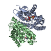 4ri6S S: Starting model for refinement C: citing same article ( |
|---|---|
| Similar structure data |


-Links
 PDBj
PDBj

- Assembly
| Deposited unit | 
| ||||||||
|---|---|---|---|---|---|---|---|---|---|
| 1 | 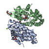
| ||||||||
| 2 | 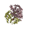
| ||||||||
| Unit cell |
|
-Components
-Protein , 1 types, 4 molecules ABCD
| #1: Protein | Mass: 23091.201 Da / Num. of mol.: 4 Source method: isolated from a genetically manipulated source Source: (gene. exp.)  |
|---|
-Non-polymers , 7 types, 889 molecules 


| #2: Chemical | ChemComp-GSH /  Mass: 307.323 Da / Num. of mol.: 4 / Source method: obtained synthetically / Formula: C10H17N3O6S Mass: 307.323 Da / Num. of mol.: 4 / Source method: obtained synthetically / Formula: C10H17N3O6S#3: Chemical | ChemComp-GOL /  Mass: 92.094 Da / Num. of mol.: 4 / Source method: obtained synthetically / Formula: C3H8O3 Mass: 92.094 Da / Num. of mol.: 4 / Source method: obtained synthetically / Formula: C3H8O3#4: Chemical |  Mass: 24.305 Da / Num. of mol.: 2 / Source method: obtained synthetically / Formula: Mg Mass: 24.305 Da / Num. of mol.: 2 / Source method: obtained synthetically / Formula: Mg#5: Chemical |  Mass: 546.646 Da / Num. of mol.: 2 / Source method: obtained synthetically / Formula: C24H50O13 / Comment: precipitant*YM Mass: 546.646 Da / Num. of mol.: 2 / Source method: obtained synthetically / Formula: C24H50O13 / Comment: precipitant*YM#6: Chemical | ChemComp-PG4 / |  Mass: 194.226 Da / Num. of mol.: 1 / Source method: obtained synthetically / Formula: C8H18O5 / Comment: precipitant*YM Mass: 194.226 Da / Num. of mol.: 1 / Source method: obtained synthetically / Formula: C8H18O5 / Comment: precipitant*YM#7: Chemical | ChemComp-PGE / |  Mass: 150.173 Da / Num. of mol.: 1 / Source method: obtained synthetically / Formula: C6H14O4 Mass: 150.173 Da / Num. of mol.: 1 / Source method: obtained synthetically / Formula: C6H14O4#8: Water | ChemComp-HOH / | Mass: 18.015 Da / Num. of mol.: 875 / Source method: isolated from a natural source / Formula: H2O |
|---|
-Experimental details
-Experiment
| Experiment | Method: X-RAY DIFFRACTION |
|---|
- Sample preparation
| Crystal | Density Matthews: 2.22 Å3/Da / Density % sol: 44.63 % |
|---|---|
| Crystal grow | Temperature: 277 K / Method: microbatch / pH: 8.5 / Details: 30% PEG 4000, Tris HCl, 0.2 M Mg chloride |
-Data collection
| Diffraction | Mean temperature: 100 K |
|---|---|
| Diffraction source | Source: SYNCHROTRON / Site: ESRF / Beamline: BM30A / Wavelength: 0.979961 Å |
| Detector | Type: ADSC QUANTUM 315r / Detector: CCD / Date: Apr 12, 2014 |
| Radiation | Protocol: SINGLE WAVELENGTH / Monochromatic (M) / Laue (L): M / Scattering type: x-ray |
| Radiation wavelength | Wavelength: 0.979961 Å / Relative weight: 1 |
| Reflection | Resolution: 1.7→49.06 Å / Num. obs: 88400 / % possible obs: 99.6 % / Redundancy: 3.8 % / Rmerge(I) obs: 0.102 / Net I/σ(I): 11 |
| Reflection shell | Resolution: 1.7→1.79 Å / Redundancy: 3.7 % / Rmerge(I) obs: 0.565 / Mean I/σ(I) obs: 3 / % possible all: 92 |
- Processing
| Software |
| |||||||||||||||||||||||||||||||||||||||||||||||||||||||||||||||||||||||||||||||||||||||||||||||||||||||||||||||||||||||||||||||||||||||||||||||||||||||||||||||||||||||||||||||||||||||||||||||||||||||||||||||||||||||||
|---|---|---|---|---|---|---|---|---|---|---|---|---|---|---|---|---|---|---|---|---|---|---|---|---|---|---|---|---|---|---|---|---|---|---|---|---|---|---|---|---|---|---|---|---|---|---|---|---|---|---|---|---|---|---|---|---|---|---|---|---|---|---|---|---|---|---|---|---|---|---|---|---|---|---|---|---|---|---|---|---|---|---|---|---|---|---|---|---|---|---|---|---|---|---|---|---|---|---|---|---|---|---|---|---|---|---|---|---|---|---|---|---|---|---|---|---|---|---|---|---|---|---|---|---|---|---|---|---|---|---|---|---|---|---|---|---|---|---|---|---|---|---|---|---|---|---|---|---|---|---|---|---|---|---|---|---|---|---|---|---|---|---|---|---|---|---|---|---|---|---|---|---|---|---|---|---|---|---|---|---|---|---|---|---|---|---|---|---|---|---|---|---|---|---|---|---|---|---|---|---|---|---|---|---|---|---|---|---|---|---|---|---|---|---|---|---|---|---|
| Refinement | Method to determine structure: MOLECULAR REPLACEMENT Starting model: 4RI6 Resolution: 1.7→43.35 Å / SU ML: 0.15 / Cross valid method: FREE R-VALUE / σ(F): 1.35 / Phase error: 18.5 / Stereochemistry target values: ML
| |||||||||||||||||||||||||||||||||||||||||||||||||||||||||||||||||||||||||||||||||||||||||||||||||||||||||||||||||||||||||||||||||||||||||||||||||||||||||||||||||||||||||||||||||||||||||||||||||||||||||||||||||||||||||
| Solvent computation | Shrinkage radii: 0.9 Å / VDW probe radii: 1.11 Å / Solvent model: FLAT BULK SOLVENT MODEL | |||||||||||||||||||||||||||||||||||||||||||||||||||||||||||||||||||||||||||||||||||||||||||||||||||||||||||||||||||||||||||||||||||||||||||||||||||||||||||||||||||||||||||||||||||||||||||||||||||||||||||||||||||||||||
| Refinement step | Cycle: LAST / Resolution: 1.7→43.35 Å
| |||||||||||||||||||||||||||||||||||||||||||||||||||||||||||||||||||||||||||||||||||||||||||||||||||||||||||||||||||||||||||||||||||||||||||||||||||||||||||||||||||||||||||||||||||||||||||||||||||||||||||||||||||||||||
| Refine LS restraints |
| |||||||||||||||||||||||||||||||||||||||||||||||||||||||||||||||||||||||||||||||||||||||||||||||||||||||||||||||||||||||||||||||||||||||||||||||||||||||||||||||||||||||||||||||||||||||||||||||||||||||||||||||||||||||||
| LS refinement shell |
|
