 Movie
Movie Controller


+ Open data

- Basic information
| Entry | Database: PDB / ID: 5exr | |||||||||
|---|---|---|---|---|---|---|---|---|---|---|
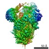
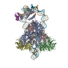
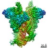
| Title | Crystal structure of human primosome | |||||||||
 Components Components |
| |||||||||
 Keywords Keywords | REPLICATION / human primosome / complex / primase / DNA polymerase alpha / primer / DNA replication / DNA / RNA / replicase | |||||||||
| Function / homology |  Function and homology information Function and homology informationribonucleotide binding / DNA primase AEP / DNA replication initiation / DNA/RNA hybrid binding / Inhibition of replication initiation of damaged DNA by RB1/E2F1 / alpha DNA polymerase:primase complex / regulation of type I interferon production / Telomere C-strand synthesis initiation / Polymerase switching / Processive synthesis on the lagging strand ...ribonucleotide binding / DNA primase AEP / DNA replication initiation / DNA/RNA hybrid binding / Inhibition of replication initiation of damaged DNA by RB1/E2F1 / alpha DNA polymerase:primase complex / regulation of type I interferon production / Telomere C-strand synthesis initiation / Polymerase switching / Processive synthesis on the lagging strand / Removal of the Flap Intermediate / lagging strand elongation / DNA replication, synthesis of primer / mitotic DNA replication initiation / Polymerase switching on the C-strand of the telomere / DNA strand elongation involved in DNA replication / leading strand elongation / G1/S-Specific Transcription / DNA synthesis involved in DNA repair / DNA replication origin binding / Activation of the pre-replicative complex / DNA replication initiation / Defective pyroptosis / double-strand break repair via nonhomologous end joining / nuclear matrix / protein import into nucleus / DNA-directed RNA polymerase activity / nuclear envelope / single-stranded DNA binding / 4 iron, 4 sulfur cluster binding / DNA-directed DNA polymerase / DNA-directed DNA polymerase activity / DNA replication / ciliary basal body / nucleotide binding / DNA repair / chromatin binding / protein kinase binding / chromatin / nucleolus / magnesium ion binding / DNA binding / zinc ion binding / nucleoplasm / membrane / metal ion binding / nucleus / cytosol Similarity search - Function | |||||||||
| Biological species |  Homo sapiens (human) Homo sapiens (human) | |||||||||
| Method |  X-RAY DIFFRACTION / SYNCHROTRON / MOLECULAR REPLACEMENT / molecular replacement / Resolution: 3.6 Å X-RAY DIFFRACTION / SYNCHROTRON / MOLECULAR REPLACEMENT / molecular replacement / Resolution: 3.6 Å | |||||||||
 Authors Authors | Tahirov, T.H. / Baranovskiy, A.G. / Babayeva, N.D. | |||||||||
| Funding support |  United States, 2items United States, 2items
| |||||||||
 Citation Citation | Journal: J.Biol.Chem. / Year: 2016 Title: Mechanism of Concerted RNA-DNA Primer Synthesis by the Human Primosome. Authors: Baranovskiy, A.G. / Babayeva, N.D. / Zhang, Y. / Gu, J. / Suwa, Y. / Pavlov, Y.I. / Tahirov, T.H. | |||||||||
| History |
|
- Structure visualization
| Structure viewer | Molecule: MolmilJmol/JSmol |
|---|
- Downloads & links
-Download
| PDBx/mmCIF format | 5exr.cif.gz | 956.6 KB | Display | PDBx/mmCIF format |
|---|---|---|---|---|
| PDB format | pdb5exr.ent.gz | 762.3 KB | Display | PDB format |
| PDBx/mmJSON format | 5exr.json.gz | Tree view | PDBx/mmJSON format | |
| Others |  Other downloads Other downloads |
-Validation report
| Arichive directory | https://data.pdbj.org/pub/pdb/validation_reports/ex/5exrftp://data.pdbj.org/pub/pdb/validation_reports/ex/5exr | HTTPS FTP |
|---|
-Related structure data
| Related structure data | 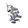 5f0qC  5f0sC  4q5vS 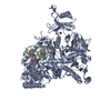 4qclS 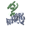 4rr2S 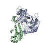 4y97S S: Starting model for refinement C: citing same article ( |
|---|---|
| Similar structure data |


-Links
 PDBj
PDBj


- Assembly
| Deposited unit | 
| ||||||||
|---|---|---|---|---|---|---|---|---|---|
| 1 | 
| ||||||||
| 2 | 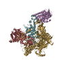
| ||||||||
| Unit cell |
| ||||||||
| Details | tetramer according to electrophoresis |
-Components
-Protein , 2 types, 4 molecules AEBF
| #1: Protein | Mass: 49981.012 Da / Num. of mol.: 2 Source method: isolated from a genetically manipulated source Source: (gene. exp.) Homo sapiens (human) / Gene: PRIM1 / Production host:  References: UniProt: P49642, Transferases; Transferring phosphorus-containing groups; Nucleotidyltransferases #2: Protein | Mass: 58890.918 Da / Num. of mol.: 2 Source method: isolated from a genetically manipulated source Source: (gene. exp.) Homo sapiens (human) / Gene: PRIM2, PRIM2A / Production host: References: UniProt: P49643, Transferases; Transferring phosphorus-containing groups; Nucleotidyltransferases |
|---|
-DNA polymerase alpha ... , 2 types, 4 molecules CGDH
| #3: Protein | Mass: 129308.773 Da / Num. of mol.: 2 / Mutation: V516A Source method: isolated from a genetically manipulated source Source: (gene. exp.) Homo sapiens (human) / Gene: POLA1, POLA / Production host:   Spodoptera frugiperda (fall armyworm) / References: UniProt: P09884, DNA-directed DNA polymerase Spodoptera frugiperda (fall armyworm) / References: UniProt: P09884, DNA-directed DNA polymerase#4: Protein | Mass: 65884.344 Da / Num. of mol.: 2 Source method: isolated from a genetically manipulated source Source: (gene. exp.) Homo sapiens (human) / Gene: POLA2 / Production host: Spodoptera frugiperda (fall armyworm) / References: UniProt: Q14181 |
|---|
-Non-polymers , 2 types, 8 molecules 

| #5: Chemical | ChemComp-ZN /  Mass: 65.409 Da / Num. of mol.: 6 / Source method: obtained synthetically / Formula: Zn Mass: 65.409 Da / Num. of mol.: 6 / Source method: obtained synthetically / Formula: Zn#6: Chemical |  Mass: 351.640 Da / Num. of mol.: 2 / Source method: obtained synthetically / Formula: Fe4S4 Mass: 351.640 Da / Num. of mol.: 2 / Source method: obtained synthetically / Formula: Fe4S4 |
|---|
-Details
| Has protein modification | Y |
|---|
-Experimental details
-Experiment
| Experiment | Method: X-RAY DIFFRACTION / Number of used crystals: 1 |
|---|
- Sample preparation
| Crystal | Density Matthews: 3.37 Å3/Da / Density % sol: 63.46 % / Description: thin plate in form of parallelogram |
|---|---|
| Crystal grow | Temperature: 295 K / Method: vapor diffusion, sitting drop / pH: 8.5 Details: 0.2 M lithium sulphate, 50 mM TRIS HCl pH 8.5, 2 mM TCEP pH 7.5, 11.2% w/v PEG 4,000, 3% v/v ethanol, 0.5% v/v polypropylene glycol P400 and 0.2 mM EDTA |
-Data collection
| Diffraction | Mean temperature: 100 K |
|---|---|
| Diffraction source | Source: SYNCHROTRON / Site: APS / Beamline: 24-ID-C / Wavelength: 0.9795 Å |
| Detector | Type: DECTRIS PILATUS 6M-F / Detector: PIXEL / Date: Mar 1, 2013 |
| Radiation | Protocol: SINGLE WAVELENGTH / Monochromatic (M) / Laue (L): M / Scattering type: x-ray |
| Radiation wavelength | Wavelength: 0.9795 Å / Relative weight: 1 |
| Reflection | Resolution: 3.6→50 Å / Num. obs: 74238 / % possible obs: 80.5 % / Observed criterion σ(I): -1 / Redundancy: 2.6 % / Rmerge(I) obs: 0.063 / Χ2: 2.353 / Net I/av σ(I): 8 / Net I/σ(I): 12.2 / Num. measured all: 236349 |
| Reflection shell | Resolution: 3.6→3.66 Å / Redundancy: 1.9 % / Rmerge(I) obs: 0.364 / Mean I/σ(I) obs: 1.93 / Num. unique all: 3348 / Χ2: 0.764 / Rejects: 0 / % possible all: 72.7 |
-Phasing
| Phasing | Method: molecular replacement |
|---|
- Processing
| Software |
| ||||||||||||||||||||||||||||||||||||
|---|---|---|---|---|---|---|---|---|---|---|---|---|---|---|---|---|---|---|---|---|---|---|---|---|---|---|---|---|---|---|---|---|---|---|---|---|---|
| Refinement | Method to determine structure: MOLECULAR REPLACEMENT Starting model: 4QCL, 4Q5V, 4RR2, 4Y97 Resolution: 3.6→39.94 Å / Data cutoff high absF: 6416908 / Data cutoff low absF: 0 / Isotropic thermal model: RESTRAINED / Cross valid method: THROUGHOUT / σ(F): 1
| ||||||||||||||||||||||||||||||||||||
| Solvent computation | Solvent model: FLAT MODEL / Bsol: 161.81 Å2 / ksol: 0.4022 e/Å3 | ||||||||||||||||||||||||||||||||||||
| Displacement parameters | Biso max: 145.71 Å2 / Biso mean: 61.2 Å2 / Biso min: 1.1 Å2
| ||||||||||||||||||||||||||||||||||||
| Refine analyze |
| ||||||||||||||||||||||||||||||||||||
| Refinement step | Cycle: final / Resolution: 3.6→39.94 Å
| ||||||||||||||||||||||||||||||||||||
| Refine LS restraints |
| ||||||||||||||||||||||||||||||||||||
| LS refinement shell | Resolution: 3.6→3.83 Å / Rfactor Rfree error: 0.02 / Total num. of bins used: 6
| ||||||||||||||||||||||||||||||||||||
| Xplor file |
|
