 Movie
Movie Controller


+ Open data

- Basic information
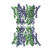
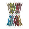
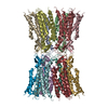
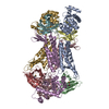
| Entry | Database: PDB / ID: 5era | ||||||
|---|---|---|---|---|---|---|---|
| Title | Human Connexin-26 (Calcium-free) | ||||||
 Components Components | Gap junction beta-2 protein | ||||||
 Keywords Keywords | CALCIUM BINDING PROTEIN / gap junction / ion channel / calcium binding / electrostatic gating | ||||||
| Function / homology |  Function and homology information Function and homology informationTransport of connexons to the plasma membrane / gap junction channel activity involved in cell communication by electrical coupling / gap junction-mediated intercellular transport / Oligomerization of connexins into connexons / Transport of connexins along the secretory pathway / gap junction assembly / connexin complex / gap junction / Gap junction assembly / gap junction channel activity ...Transport of connexons to the plasma membrane / gap junction channel activity involved in cell communication by electrical coupling / gap junction-mediated intercellular transport / Oligomerization of connexins into connexons / Transport of connexins along the secretory pathway / gap junction assembly / connexin complex / gap junction / Gap junction assembly / gap junction channel activity / endoplasmic reticulum-Golgi intermediate compartment / sensory perception of sound / transmembrane transport / cell-cell signaling / calcium ion binding / identical protein binding / plasma membrane Similarity search - Function | ||||||
| Biological species |  Homo sapiens (human) Homo sapiens (human) | ||||||
| Method |  X-RAY DIFFRACTION / SYNCHROTRON / MOLECULAR REPLACEMENT / Resolution: 3.8 Å X-RAY DIFFRACTION / SYNCHROTRON / MOLECULAR REPLACEMENT / Resolution: 3.8 Å | ||||||
 Authors Authors | Purdy, M.D. / Bennett, B.C. / Baker, K.A. / Yeager, M.J. | ||||||
 Citation Citation | Journal: Nat Commun / Year: 2016 Title: An electrostatic mechanism for Ca(2+)-mediated regulation of gap junction channels. Authors: Bennett, B.C. / Purdy, M.D. / Baker, K.A. / Acharya, C. / McIntire, W.E. / Stevens, R.C. / Zhang, Q. / Harris, A.L. / Abagyan, R. / Yeager, M. #1: Journal: Proc. Natl. Acad. Sci. U.S.A. / Year: 2013Title: Steroid-based facial amphiphiles for stabilization and crystallization of membrane proteins. Authors: Lee, S.C. / Bennett, B.C. / Hong, W.X. / Fu, Y. / Baker, K.A. / Marcoux, J. / Robinson, C.V. / Ward, A.B. / Halpert, J.R. / Stevens, R.C. / Stout, C.D. / Yeager, M.J. / Zhang, Q. | ||||||
| History |
|
- Structure visualization
| Structure viewer | Molecule: MolmilJmol/JSmol |
|---|
- Downloads & links
-Download
| PDBx/mmCIF format | 5era.cif.gz | 78.6 KB | Display | PDBx/mmCIF format |
|---|---|---|---|---|
| PDB format | pdb5era.ent.gz | 58.5 KB | Display | PDB format |
| PDBx/mmJSON format | 5era.json.gz | Tree view | PDBx/mmJSON format | |
| Others |  Other downloads Other downloads |
-Validation report
| Arichive directory | https://data.pdbj.org/pub/pdb/validation_reports/er/5eraftp://data.pdbj.org/pub/pdb/validation_reports/er/5era | HTTPS FTP |
|---|
-Related structure data
| Related structure data |  5er7SC S: Starting model for refinement C: citing same article ( |
|---|---|
| Similar structure data |
-Links
 PDBj
PDBj- Assembly
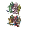
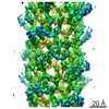
| Deposited unit | 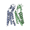
| ||||||||
|---|---|---|---|---|---|---|---|---|---|
| 1 | 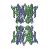
| ||||||||
| 2 | 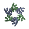
| ||||||||
| Unit cell |
| ||||||||
| Symmetry | Point symmetry: (Schoenflies symbol: D6 (2x6 fold dihedral)) |
-Components
| #1: Protein | Mass: 26138.848 Da / Num. of mol.: 2 / Fragment: full length protein / Mutation: C211S, C218S Source method: isolated from a genetically manipulated source Source: (gene. exp.) Homo sapiens (human) / Gene: GJB2 / Plasmid: pVL1393 / Production host:   Spodoptera frugiperda (fall armyworm) / Strain (production host): Sf9 / References: UniProt: P29033 Spodoptera frugiperda (fall armyworm) / Strain (production host): Sf9 / References: UniProt: P29033Has protein modification | Y | |
|---|
-Experimental details
-Experiment
| Experiment | Method: X-RAY DIFFRACTION / Number of used crystals: 1 |
|---|
- Sample preparation
| Crystal | Density Matthews: 3.59 Å3/Da / Density % sol: 65.71 % |
|---|---|
| Crystal grow | Temperature: 298 K / Method: vapor diffusion, hanging drop / pH: 7.5 Details: 0.1 M Hepes, 15 mM Sodium formate, 14% (w/v) PEG 3350 |
-Data collection
| Diffraction | Mean temperature: 100 K | ||||||||||||||||||||||||||||||||||||||||||||||||||||||||||||||||||||||||||||||||||||||||||||||||||||||||||||||||||||||||||||||
|---|---|---|---|---|---|---|---|---|---|---|---|---|---|---|---|---|---|---|---|---|---|---|---|---|---|---|---|---|---|---|---|---|---|---|---|---|---|---|---|---|---|---|---|---|---|---|---|---|---|---|---|---|---|---|---|---|---|---|---|---|---|---|---|---|---|---|---|---|---|---|---|---|---|---|---|---|---|---|---|---|---|---|---|---|---|---|---|---|---|---|---|---|---|---|---|---|---|---|---|---|---|---|---|---|---|---|---|---|---|---|---|---|---|---|---|---|---|---|---|---|---|---|---|---|---|---|---|
| Diffraction source | Source: SYNCHROTRON / Site: APS  / Beamline: 23-ID-D / Wavelength: 1 Å / Beamline: 23-ID-D / Wavelength: 1 Å | ||||||||||||||||||||||||||||||||||||||||||||||||||||||||||||||||||||||||||||||||||||||||||||||||||||||||||||||||||||||||||||||
| Detector | Type: MARMOSAIC 300 mm CCD / Detector: CCD / Date: Jun 15, 2009 | ||||||||||||||||||||||||||||||||||||||||||||||||||||||||||||||||||||||||||||||||||||||||||||||||||||||||||||||||||||||||||||||
| Radiation | Protocol: SINGLE WAVELENGTH / Monochromatic (M) / Laue (L): M / Scattering type: x-ray | ||||||||||||||||||||||||||||||||||||||||||||||||||||||||||||||||||||||||||||||||||||||||||||||||||||||||||||||||||||||||||||||
| Radiation wavelength | Wavelength: 1 Å / Relative weight: 1 | ||||||||||||||||||||||||||||||||||||||||||||||||||||||||||||||||||||||||||||||||||||||||||||||||||||||||||||||||||||||||||||||
| Reflection | Resolution: 3.3→50 Å / Num. obs: 7398 / % possible obs: 99.9 % / Redundancy: 7.4 % / Biso Wilson estimate: 152.13 Å2 / Rmerge(I) obs: 0.082 / Χ2: 1.184 / Net I/av σ(I): 21.375 / Net I/σ(I): 5.9 / Num. measured all: 85071 | ||||||||||||||||||||||||||||||||||||||||||||||||||||||||||||||||||||||||||||||||||||||||||||||||||||||||||||||||||||||||||||||
| Reflection shell | Diffraction-ID: 1 / Rejects: _
|
- Processing
| Software |
| ||||||||||||||||||||||||||||||||||||||||||
|---|---|---|---|---|---|---|---|---|---|---|---|---|---|---|---|---|---|---|---|---|---|---|---|---|---|---|---|---|---|---|---|---|---|---|---|---|---|---|---|---|---|---|---|
| Refinement | Method to determine structure: MOLECULAR REPLACEMENT Starting model: 5ER7 Resolution: 3.8→44.132 Å / SU ML: 0.55 / Cross valid method: FREE R-VALUE / σ(F): 1.35 / Phase error: 40.35 / Stereochemistry target values: ML
| ||||||||||||||||||||||||||||||||||||||||||
| Solvent computation | Shrinkage radii: 0.9 Å / VDW probe radii: 1.11 Å / Solvent model: FLAT BULK SOLVENT MODEL | ||||||||||||||||||||||||||||||||||||||||||
| Displacement parameters | Biso max: 207.98 Å2 / Biso mean: 127.161 Å2 / Biso min: 58.25 Å2 | ||||||||||||||||||||||||||||||||||||||||||
| Refinement step | Cycle: final / Resolution: 3.8→44.132 Å
| ||||||||||||||||||||||||||||||||||||||||||
| Refine LS restraints |
| ||||||||||||||||||||||||||||||||||||||||||
| LS refinement shell | Refine-ID: X-RAY DIFFRACTION / Total num. of bins used: 5
|
