 Movie
Movie Controller

[English] 日本語
 Yorodumi
- PDB-5b2g: Crystal structure of human claudin-4 in complex with C-terminal f... -
+ Open data

- Basic information
| Entry | Database: PDB / ID: 5b2g | ||||||||||||
|---|---|---|---|---|---|---|---|---|---|---|---|---|---|
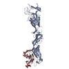
| Title | Crystal structure of human claudin-4 in complex with C-terminal fragment of Clostridium perfringens enterotoxin | ||||||||||||
 Components Components |
| ||||||||||||
 Keywords Keywords | MEMBRANE PROTEIN / Complex / Cell-free protein expression system | ||||||||||||
| Function / homology |  Function and homology information Function and homology informationparacellular transport / calcium-independent cell-cell adhesion / Tight junction interactions / apicolateral plasma membrane / bicellular tight junction assembly / regulation of cell morphogenesis / tight junction / positive regulation of wound healing / renal absorption / chloride channel activity ...paracellular transport / calcium-independent cell-cell adhesion / Tight junction interactions / apicolateral plasma membrane / bicellular tight junction assembly / regulation of cell morphogenesis / tight junction / positive regulation of wound healing / renal absorption / chloride channel activity / establishment of skin barrier / lateral plasma membrane / bicellular tight junction / chloride channel complex / viral release from host cell by cytolysis / peptidoglycan catabolic process / response to progesterone / basal plasma membrane / female pregnancy / circadian rhythm / cell wall macromolecule catabolic process / lysozyme / cell-cell junction / lysozyme activity / transmembrane signaling receptor activity / toxin activity / host cell cytoplasm / cell adhesion / defense response to bacterium / apical plasma membrane / positive regulation of cell migration / structural molecule activity / extracellular region / identical protein binding / plasma membrane Similarity search - Function | ||||||||||||
| Biological species |  Enterobacteria phage T4 (virus) Enterobacteria phage T4 (virus) Homo sapiens (human) Homo sapiens (human)  Clostridium perfringens (bacteria) Clostridium perfringens (bacteria) | ||||||||||||
| Method |  X-RAY DIFFRACTION / SYNCHROTRON / MOLECULAR REPLACEMENT / Resolution: 3.5 Å X-RAY DIFFRACTION / SYNCHROTRON / MOLECULAR REPLACEMENT / Resolution: 3.5 Å | ||||||||||||
 Authors Authors | Shinoda, T. / Kimura-Someya, T. / Shirouzu, M. / Yokoyama, S. | ||||||||||||
| Funding support |  Japan, 3items Japan, 3items
| ||||||||||||
 Citation Citation | Journal: Sci Rep / Year: 2016 Title: Structural basis for disruption of claudin assembly in tight junctions by an enterotoxin Authors: Shinoda, T. / Shinya, N. / Ito, K. / Ohsawa, N. / Terada, T. / Hirata, K. / Kawano, Y. / Yamamoto, M. / Kimura-Someya, T. / Yokoyama, S. / Shirouzu, M. | ||||||||||||
| History |
|
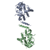
- Structure visualization
| Structure viewer | Molecule: MolmilJmol/JSmol |
|---|
- Downloads & links
-Download
| PDBx/mmCIF format | 5b2g.cif.gz | 720.9 KB | Display | PDBx/mmCIF format |
|---|---|---|---|---|
| PDB format | pdb5b2g.ent.gz | 599 KB | Display | PDB format |
| PDBx/mmJSON format | 5b2g.json.gz | Tree view | PDBx/mmJSON format | |
| Others |  Other downloads Other downloads |
-Validation report
| Arichive directory | https://data.pdbj.org/pub/pdb/validation_reports/b2/5b2gftp://data.pdbj.org/pub/pdb/validation_reports/b2/5b2g | HTTPS FTP |
|---|
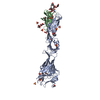
-Related structure data
| Related structure data |  3am2S S: Starting model for refinement |
|---|---|
| Similar structure data |


-Links
 PDBj
PDBj


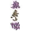
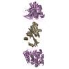
- Assembly
| Deposited unit | 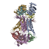
| ||||||||
|---|---|---|---|---|---|---|---|---|---|
| 1 | 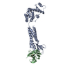
| ||||||||
| 2 | 
| ||||||||
| 3 | 
| ||||||||
| 4 | 
| ||||||||
| Unit cell |
|
-Components
| #1: Protein | Mass: 39410.793 Da / Num. of mol.: 4 / Fragment: UNP residues 2-162,UNP residues 1-183 / Mutation: R1012G, C1054T, C1097A, I1137R Source method: isolated from a genetically manipulated source Source: (gene. exp.) Enterobacteria phage T4 (virus), (gene. exp.) Homo sapiens (human)Gene: CLDN4, CPER, CPETR1, WBSCR8 Details (production host): Sample was prepared by the E.coli cell-free protein synthesis system Production host: #2: Protein | Mass: 15879.505 Da / Num. of mol.: 4 / Fragment: UNP residues 187-319 Source method: isolated from a genetically manipulated source Source: (gene. exp.) Clostridium perfringens (bacteria) / Gene: cpeDetails (production host): Sample was prepared by the E.coli cell-free protein synthesis system Production host: Has protein modification | Y | |
|---|
-Experimental details
-Experiment
| Experiment | Method: X-RAY DIFFRACTION / Number of used crystals: 1 |
|---|
- Sample preparation
| Crystal | Density Matthews: 3.33 Å3/Da / Density % sol: 63.07 % Description: THE ENTRY CONTAINS FRIEDEL PAIRS IN F_PLUS/MINUS COLUMNS. |
|---|---|
| Crystal grow | Temperature: 288 K / Method: vapor diffusion, hanging drop Details: 75mM MES-NaOH, 20% PEG3350, 7-10% 1,6-hexanediol, 0.002% NaN3, 0.0005% 2,6-di-t-butyl-p-cresol, 150mM NaCl PH range: 5.0 - 5.5 |
-Data collection
| Diffraction | Mean temperature: 100 K |
|---|---|
| Diffraction source | Source: SYNCHROTRON / Site: SPring-8 / Beamline: BL41XU / Wavelength: 0.9793 Å |
| Detector | Type: RAYONIX MX225HE / Detector: CCD / Date: Dec 13, 2013 |
| Radiation | Protocol: SINGLE WAVELENGTH / Monochromatic (M) / Laue (L): M / Scattering type: x-ray |
| Radiation wavelength | Wavelength: 0.9793 Å / Relative weight: 1 |
| Reflection | Resolution: 3.35→50 Å / Num. obs: 76023 / % possible obs: 99.6 % / Redundancy: 4.56 % / Rmerge(I) obs: 0.161 / Net I/σ(I): 6.63 |
- Processing
| Software |
| |||||||||||||||||||||||||||||||||||||||||||||||||||||||||||||||||||||||||||||||||||||||||||
|---|---|---|---|---|---|---|---|---|---|---|---|---|---|---|---|---|---|---|---|---|---|---|---|---|---|---|---|---|---|---|---|---|---|---|---|---|---|---|---|---|---|---|---|---|---|---|---|---|---|---|---|---|---|---|---|---|---|---|---|---|---|---|---|---|---|---|---|---|---|---|---|---|---|---|---|---|---|---|---|---|---|---|---|---|---|---|---|---|---|---|---|---|
| Refinement | Method to determine structure: MOLECULAR REPLACEMENT Starting model: 3AM2 Resolution: 3.5→48.589 Å / SU ML: 0.8 / Cross valid method: FREE R-VALUE / σ(F): 1.92 / Phase error: 34.28 / Stereochemistry target values: MLHL
| |||||||||||||||||||||||||||||||||||||||||||||||||||||||||||||||||||||||||||||||||||||||||||
| Solvent computation | Shrinkage radii: 0.9 Å / VDW probe radii: 1.11 Å / Solvent model: FLAT BULK SOLVENT MODEL | |||||||||||||||||||||||||||||||||||||||||||||||||||||||||||||||||||||||||||||||||||||||||||
| Refinement step | Cycle: LAST / Resolution: 3.5→48.589 Å
| |||||||||||||||||||||||||||||||||||||||||||||||||||||||||||||||||||||||||||||||||||||||||||
| Refine LS restraints |
| |||||||||||||||||||||||||||||||||||||||||||||||||||||||||||||||||||||||||||||||||||||||||||
| LS refinement shell |
| |||||||||||||||||||||||||||||||||||||||||||||||||||||||||||||||||||||||||||||||||||||||||||
| Refinement TLS params. | Method: refined / Origin x: 40.7097 Å / Origin y: -12.0877 Å / Origin z: -5.8343 Å
| |||||||||||||||||||||||||||||||||||||||||||||||||||||||||||||||||||||||||||||||||||||||||||
| Refinement TLS group | Selection details: all |
