 Movie
Movie Controller

[English] 日本語
 Yorodumi
- PDB-4e8u: Crystal structure of Arabidopsis IDN2 XS domain along with a smal... -
+ Open data

- Basic information
| Entry | Database: PDB / ID: 4e8u | ||||||
|---|---|---|---|---|---|---|---|
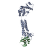
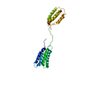
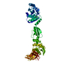
| Title | Crystal structure of Arabidopsis IDN2 XS domain along with a small segment of adjacent coiled-coil region | ||||||
 Components Components | Putative uncharacterized protein T8P19.180 | ||||||
 Keywords Keywords | RNA BINDING PROTEIN / XS domain / RNA directed DNA methylation / RNA | ||||||
| Function / homology |  Function and homology information Function and homology informationgene silencing by siRNA-directed DNA methylation / regulation of double-strand break repair via homologous recombination / response to cadmium ion / RNA binding Similarity search - Function | ||||||
| Biological species |  | ||||||
| Method |  X-RAY DIFFRACTION / SYNCHROTRON / SAD (using a smaller construct), molecular replacement / Resolution: 2.701 Å X-RAY DIFFRACTION / SYNCHROTRON / SAD (using a smaller construct), molecular replacement / Resolution: 2.701 Å | ||||||
 Authors Authors | Simanshu, D.K. / Patel, D.J. | ||||||
 Citation Citation | Journal: Proc.Natl.Acad.Sci.USA / Year: 2012 Title: INVOLVED IN DE NOVO 2-containing complex involved in RNA-directed DNA methylation in Arabidopsis. Authors: Ausin, I. / Greenberg, M.V. / Simanshu, D.K. / Hale, C.J. / Vashisht, A.A. / Simon, S.A. / Lee, T.F. / Feng, S. / Espanola, S.D. / Meyers, B.C. / Wohlschlegel, J.A. / Patel, D.J. / Jacobsen, S.E. | ||||||
| History |
|
- Structure visualization
| Structure viewer | Molecule: MolmilJmol/JSmol |
|---|
- Downloads & links
-Download
| PDBx/mmCIF format | 4e8u.cif.gz | 146.4 KB | Display | PDBx/mmCIF format |
|---|---|---|---|---|
| PDB format | pdb4e8u.ent.gz | 117.3 KB | Display | PDB format |
| PDBx/mmJSON format | 4e8u.json.gz | Tree view | PDBx/mmJSON format | |
| Others |  Other downloads Other downloads |
-Validation report
| Arichive directory | https://data.pdbj.org/pub/pdb/validation_reports/e8/4e8uftp://data.pdbj.org/pub/pdb/validation_reports/e8/4e8u | HTTPS FTP |
|---|
-Related structure data
| Similar structure data |
|---|


-Links
 PDBj
PDBj- Assembly
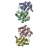
| Deposited unit | 
| |||||||||||||||||||||||||||
|---|---|---|---|---|---|---|---|---|---|---|---|---|---|---|---|---|---|---|---|---|---|---|---|---|---|---|---|---|
| 1 |
| |||||||||||||||||||||||||||
| 2 | 
| |||||||||||||||||||||||||||
| Unit cell |
| |||||||||||||||||||||||||||
| Components on special symmetry positions |
| |||||||||||||||||||||||||||
| Noncrystallographic symmetry (NCS) | NCS domain:
NCS domain segments:
NCS oper: (Code: given Matrix: (0.999664, -0.003911, -0.02561), Vector: |
-Components
| #1: Protein | Mass: 19632.113 Da / Num. of mol.: 2 Source method: isolated from a genetically manipulated source Source: (gene. exp.)  #2: Chemical | ChemComp-SO4 /   Mass: 96.063 Da / Num. of mol.: 4 / Source method: obtained synthetically / Formula: SO4 Mass: 96.063 Da / Num. of mol.: 4 / Source method: obtained synthetically / Formula: SO4#3: Water | ChemComp-HOH / |  Mass: 18.015 Da / Num. of mol.: 25 / Source method: isolated from a natural source / Formula: H2O Mass: 18.015 Da / Num. of mol.: 25 / Source method: isolated from a natural source / Formula: H2O |
|---|
-Experimental details
-Experiment
| Experiment | Method: X-RAY DIFFRACTION / Number of used crystals: 1 |
|---|
- Sample preparation
| Crystal | Density Matthews: 4.13 Å3/Da / Density % sol: 70.21 % |
|---|---|
| Crystal grow | Temperature: 293 K / Method: vapor diffusion, hanging drop / pH: 6.5 Details: 0.1 M BIS-TRIS pH 6.5, 2.0 M ammonium sulfate, VAPOR DIFFUSION, HANGING DROP, temperature 293K |
-Data collection
| Diffraction | Mean temperature: 100 K | |||||||||||||||||||||||||||||||||||||||||||||||||||||||||||||||||||||||||||||
|---|---|---|---|---|---|---|---|---|---|---|---|---|---|---|---|---|---|---|---|---|---|---|---|---|---|---|---|---|---|---|---|---|---|---|---|---|---|---|---|---|---|---|---|---|---|---|---|---|---|---|---|---|---|---|---|---|---|---|---|---|---|---|---|---|---|---|---|---|---|---|---|---|---|---|---|---|---|---|
| Diffraction source | Source: SYNCHROTRON / Site: APS  / Beamline: 24-ID-C / Wavelength: 0.9792 Å / Beamline: 24-ID-C / Wavelength: 0.9792 Å | |||||||||||||||||||||||||||||||||||||||||||||||||||||||||||||||||||||||||||||
| Detector | Type: ADSC QUANTUM 315 / Detector: CCD / Date: Mar 4, 2010 Details: Cryogenically-cooled double crystal Si(111) monochromator. Triple striped vertical and horizantal focussing mirrors in Kirkpatrick-Baez geometry | |||||||||||||||||||||||||||||||||||||||||||||||||||||||||||||||||||||||||||||
| Radiation | Monochromator: Cryo-Cooled Si(111) double crystal. / Protocol: SINGLE WAVELENGTH / Monochromatic (M) / Laue (L): M / Scattering type: x-ray | |||||||||||||||||||||||||||||||||||||||||||||||||||||||||||||||||||||||||||||
| Radiation wavelength | Wavelength: 0.9792 Å / Relative weight: 1 | |||||||||||||||||||||||||||||||||||||||||||||||||||||||||||||||||||||||||||||
| Reflection | Resolution: 2.7→50 Å / Num. obs: 18040 / % possible obs: 99 % / Redundancy: 10.3 % / Rmerge(I) obs: 0.087 / Χ2: 2.453 / Net I/σ(I): 11.2 | |||||||||||||||||||||||||||||||||||||||||||||||||||||||||||||||||||||||||||||
| Reflection shell |
|
- Processing
| Software |
| |||||||||||||||||||||||||||||||||||||||||||||||||
|---|---|---|---|---|---|---|---|---|---|---|---|---|---|---|---|---|---|---|---|---|---|---|---|---|---|---|---|---|---|---|---|---|---|---|---|---|---|---|---|---|---|---|---|---|---|---|---|---|---|---|
| Refinement | Method to determine structure: SAD (using a smaller construct), molecular replacement Resolution: 2.701→29.888 Å / Occupancy max: 1 / Occupancy min: 0.42 / SU ML: 0.36 / Cross valid method: R-Free / σ(F): 0.1 / Phase error: 37.58 / Stereochemistry target values: ML
| |||||||||||||||||||||||||||||||||||||||||||||||||
| Solvent computation | Shrinkage radii: 0.9 Å / VDW probe radii: 1.11 Å / Solvent model: FLAT BULK SOLVENT MODEL / Bsol: 48.948 Å2 / ksol: 0.302 e/Å3 | |||||||||||||||||||||||||||||||||||||||||||||||||
| Displacement parameters | Biso max: 169.8 Å2 / Biso mean: 82.6751 Å2 / Biso min: 18.43 Å2
| |||||||||||||||||||||||||||||||||||||||||||||||||
| Refinement step | Cycle: LAST / Resolution: 2.701→29.888 Å
| |||||||||||||||||||||||||||||||||||||||||||||||||
| Refine LS restraints |
| |||||||||||||||||||||||||||||||||||||||||||||||||
| Refine LS restraints NCS |
| |||||||||||||||||||||||||||||||||||||||||||||||||
| LS refinement shell | Refine-ID: X-RAY DIFFRACTION / Total num. of bins used: 6
| |||||||||||||||||||||||||||||||||||||||||||||||||
| Refinement TLS params. | Method: refined / Origin x: 16.4628 Å / Origin y: 23.7646 Å / Origin z: 72.2328 Å
| |||||||||||||||||||||||||||||||||||||||||||||||||
| Refinement TLS group |
|
