 Movie
Movie Controller

[English] 日本語
 Yorodumi
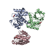
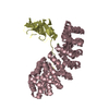
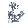
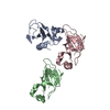
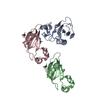
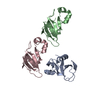
- PDB-5emo: Structure of the star domain of T-STAR in complex with AUUAAA RNA -
+ Open data

- Basic information
| Entry | Database: PDB / ID: 5emo | ||||||
|---|---|---|---|---|---|---|---|
| Title | Structure of the star domain of T-STAR in complex with AUUAAA RNA | ||||||
 Components Components |
| ||||||
 Keywords Keywords | RNA BINDING PROTEIN / Protein - RNA complexes STAR protein Alternative splicing KH domain | ||||||
| Function / homology |  Function and homology information Function and homology informationPTK6 Regulates Proteins Involved in RNA Processing / regulation of alternative mRNA splicing, via spliceosome / SH3 domain binding / mRNA processing / protein domain specific binding / mRNA binding / protein-containing complex / RNA binding / nucleoplasm / identical protein binding / nucleus Similarity search - Function | ||||||
| Biological species |  Homo sapiens (human) Homo sapiens (human)synthetic construct (others) | ||||||
| Method |  X-RAY DIFFRACTION / SYNCHROTRON / MOLECULAR REPLACEMENT / molecular replacement / Resolution: 3.03 Å X-RAY DIFFRACTION / SYNCHROTRON / MOLECULAR REPLACEMENT / molecular replacement / Resolution: 3.03 Å | ||||||
 Authors Authors | Dominguez, C. / Feracci, M. | ||||||
| Funding support |  United Kingdom, 1items United Kingdom, 1items
| ||||||
 Citation Citation | Journal: Nat Commun / Year: 2016 Title: Structural basis of RNA recognition and dimerization by the STAR proteins T-STAR and Sam68. Authors: Feracci, M. / Foot, J.N. / Grellscheid, S.N. / Danilenko, M. / Stehle, R. / Gonchar, O. / Kang, H.S. / Dalgliesh, C. / Meyer, N.H. / Liu, Y. / Lahat, A. / Sattler, M. / Eperon, I.C. / ...Authors: Feracci, M. / Foot, J.N. / Grellscheid, S.N. / Danilenko, M. / Stehle, R. / Gonchar, O. / Kang, H.S. / Dalgliesh, C. / Meyer, N.H. / Liu, Y. / Lahat, A. / Sattler, M. / Eperon, I.C. / Elliott, D.J. / Dominguez, C. | ||||||
| History |
|
- Structure visualization
| Structure viewer | Molecule: MolmilJmol/JSmol |
|---|
- Downloads & links
-Download
| PDBx/mmCIF format | 5emo.cif.gz | 77.2 KB | Display | PDBx/mmCIF format |
|---|---|---|---|---|
| PDB format | pdb5emo.ent.gz | 57.9 KB | Display | PDB format |
| PDBx/mmJSON format | 5emo.json.gz | Tree view | PDBx/mmJSON format | |
| Others |  Other downloads Other downloads |
-Validation report
| Arichive directory | https://data.pdbj.org/pub/pdb/validation_reports/em/5emoftp://data.pdbj.org/pub/pdb/validation_reports/em/5emo | HTTPS FTP |
|---|
-Related structure data
| Related structure data | 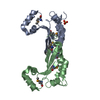 5el3SC 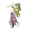 5elrC  5elsC  5eltC S: Starting model for refinement C: citing same article ( |
|---|---|
| Similar structure data |


-Links
 PDBj
PDBj
- Assembly
| Deposited unit | 
| ||||||||
|---|---|---|---|---|---|---|---|---|---|
| 1 |
| ||||||||
| Unit cell |
|
-Components
| #1: Protein | Mass: 21156.258 Da / Num. of mol.: 2 / Fragment: RNA binding protein Source method: isolated from a genetically manipulated source Details: N-terminus GA residues from tag. / Source: (gene. exp.) Homo sapiens (human) / Gene: KHDRBS3, SALP, SLM2 / Plasmid: pLEICS 03 / Production host:  #2: RNA chain | Mass: 1884.197 Da / Num. of mol.: 2 / Source method: obtained synthetically / Source: (synth.) synthetic construct (others) |
|---|
-Experimental details
-Experiment
| Experiment | Method: X-RAY DIFFRACTION / Number of used crystals: 1 |
|---|
- Sample preparation
| Crystal | Density Matthews: 2.37 Å3/Da / Density % sol: 48 % |
|---|---|
| Crystal grow | Temperature: 277 K / Method: vapor diffusion, sitting drop / pH: 7.5 / Details: NaCl, Na-HEPES, PEG 4000 |
-Data collection
| Diffraction | Mean temperature: 100 K | |||||||||||||||||||||||||||
|---|---|---|---|---|---|---|---|---|---|---|---|---|---|---|---|---|---|---|---|---|---|---|---|---|---|---|---|---|
| Diffraction source | Source: SYNCHROTRON / Site: Diamond / Beamline: I03 / Wavelength: 0.97934 Å | |||||||||||||||||||||||||||
| Detector | Type: DECTRIS PILATUS3 6M / Detector: PIXEL / Date: Feb 10, 2014 | |||||||||||||||||||||||||||
| Radiation | Protocol: SINGLE WAVELENGTH / Monochromatic (M) / Laue (L): M / Scattering type: x-ray | |||||||||||||||||||||||||||
| Radiation wavelength | Wavelength: 0.97934 Å / Relative weight: 1 | |||||||||||||||||||||||||||
| Reflection | Resolution: 3.03→28.51 Å / Num. obs: 7908 / % possible obs: 94.8 % / Redundancy: 2.6 % / Biso Wilson estimate: 55.46 Å2 / CC1/2: 0.988 / Rmerge(I) obs: 0.114 / Rpim(I) all: 0.081 / Net I/σ(I): 8.1 / Num. measured all: 20367 | |||||||||||||||||||||||||||
| Reflection shell | Diffraction-ID: 1 / Rejects: _
|
-Phasing
| Phasing | Method: molecular replacement |
|---|
- Processing
| Software |
| |||||||||||||||||||||||||||||||||||||||||||||||||
|---|---|---|---|---|---|---|---|---|---|---|---|---|---|---|---|---|---|---|---|---|---|---|---|---|---|---|---|---|---|---|---|---|---|---|---|---|---|---|---|---|---|---|---|---|---|---|---|---|---|---|
| Refinement | Method to determine structure: MOLECULAR REPLACEMENT Starting model: 5EL3 Resolution: 3.03→28.508 Å / SU ML: 0.44 / Cross valid method: THROUGHOUT / σ(F): 1.35 / Phase error: 28.53 / Stereochemistry target values: ML
| |||||||||||||||||||||||||||||||||||||||||||||||||
| Solvent computation | Shrinkage radii: 0.9 Å / VDW probe radii: 1.11 Å / Solvent model: FLAT BULK SOLVENT MODEL | |||||||||||||||||||||||||||||||||||||||||||||||||
| Displacement parameters | Biso max: 135.05 Å2 / Biso mean: 55.8537 Å2 / Biso min: 15.42 Å2 | |||||||||||||||||||||||||||||||||||||||||||||||||
| Refinement step | Cycle: final / Resolution: 3.03→28.508 Å
| |||||||||||||||||||||||||||||||||||||||||||||||||
| Refine LS restraints |
| |||||||||||||||||||||||||||||||||||||||||||||||||
| LS refinement shell | Refine-ID: X-RAY DIFFRACTION / Total num. of bins used: 6
|
