 Movie
Movie Controller

[English] 日本語
 Yorodumi
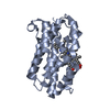
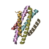
- PDB-5ehb: A de novo designed hexameric coiled-coil peptide with iodotyrosine -
+ Open data

- Basic information
| Entry | Database: PDB / ID: 5ehb | ||||||
|---|---|---|---|---|---|---|---|
| Title | A de novo designed hexameric coiled-coil peptide with iodotyrosine | ||||||
 Components Components | pHiosYI | ||||||
 Keywords Keywords | DE NOVO PROTEIN / coiled-coil / peptide design | ||||||
| Biological species | synthetic construct (others) | ||||||
| Method |  X-RAY DIFFRACTION / SYNCHROTRON / AB INITIO PHASING / Resolution: 3.19 Å X-RAY DIFFRACTION / SYNCHROTRON / AB INITIO PHASING / Resolution: 3.19 Å | ||||||
 Authors Authors | Lizatovic, R. / Aurelius, O. / Stenstrom, O. / Drakenberg, T. / Akke, M. / Logan, D.T. / Andre, I. | ||||||
 Citation Citation | Journal: Structure / Year: 2016 Title: A De Novo Designed Coiled-Coil Peptide with a Reversible pH-Induced Oligomerization Switch. Authors: Lizatovic, R. / Aurelius, O. / Stenstrom, O. / Drakenberg, T. / Akke, M. / Logan, D.T. / Andre, I. | ||||||
| History |
|
- Structure visualization
| Structure viewer | Molecule:  MolmilJmol/JSmol MolmilJmol/JSmol |
|---|
- Downloads & links
-Download
| PDBx/mmCIF format | 5ehb.cif.gz | 20.9 KB | Display | PDBx/mmCIF format |
|---|---|---|---|---|
| PDB format | pdb5ehb.ent.gz | 13.7 KB | Display | PDB format |
| PDBx/mmJSON format | 5ehb.json.gz | Tree view | PDBx/mmJSON format | |
| Others |  Other downloads Other downloads |
-Validation report
| Arichive directory | https://data.pdbj.org/pub/pdb/validation_reports/eh/5ehbftp://data.pdbj.org/pub/pdb/validation_reports/eh/5ehb | HTTPS FTP |
|---|
-Related structure data
| Similar structure data |
|---|


-Links
 PDBj
PDBj
- Assembly
| Deposited unit | 
| ||||||||||||||||||
|---|---|---|---|---|---|---|---|---|---|---|---|---|---|---|---|---|---|---|---|
| 1 | 
| ||||||||||||||||||
| Unit cell |
| ||||||||||||||||||
| Symmetry | Point symmetry: (Schoenflies symbol: C3 (3 fold cyclic)) | ||||||||||||||||||
| Noncrystallographic symmetry (NCS) | NCS domain:
NCS domain segments: Component-ID: _ / Ens-ID: 1 / Beg auth comp-ID: THR / Beg label comp-ID: THR / End auth comp-ID: ALA / End label comp-ID: ALA / Refine code: _ / Auth seq-ID: 1 - 28 / Label seq-ID: 1 - 28
|
-Components
| #1: Protein/peptide | Mass: 3853.132 Da / Num. of mol.: 2 / Mutation: Y5(IYR) / Source method: obtained synthetically / Details: Designed sequence / Source: (synth.) synthetic construct (others) Has protein modification | Y | |
|---|
-Experimental details
-Experiment
| Experiment | Method: X-RAY DIFFRACTION / Number of used crystals: 1 |
|---|
- Sample preparation
| Crystal | Density Matthews: 2.69 Å3/Da / Density % sol: 54.35 % |
|---|---|
| Crystal grow | Temperature: 293 K / Method: microbatch / pH: 7.5 Details: 2 uL 10 mg/mL pHiosYI mixed with a 2 uL solution of the below components 0.1 M Na HEPES pH 7.5 0.2 M MgCl2 10% (v/v) PEG400 Al's oil was used to separate the external reservoir, which ...Details: 2 uL 10 mg/mL pHiosYI mixed with a 2 uL solution of the below components 0.1 M Na HEPES pH 7.5 0.2 M MgCl2 10% (v/v) PEG400 Al's oil was used to separate the external reservoir, which consisted of 15% (v/v) 2-propanol. Temp details: The crystal was moved to 277 K during fishing and cryo-cooling. |
-Data collection
| Diffraction | Mean temperature: 100 K |
|---|---|
| Diffraction source | Source: SYNCHROTRON / Site: MAX II  / Beamline: I911-3 / Wavelength: 1.8 Å / Beamline: I911-3 / Wavelength: 1.8 Å |
| Detector | Type: MARMOSAIC 225 mm CCD / Detector: CCD / Date: Jan 25, 2013 |
| Radiation | Monochromator: double-crystal / Protocol: SINGLE WAVELENGTH / Monochromatic (M) / Laue (L): M / Scattering type: x-ray |
| Radiation wavelength | Wavelength: 1.8 Å / Relative weight: 1 |
| Reflection | Resolution: 3.19→27.4 Å / Num. obs: 2301 / % possible obs: 95.6 % / Redundancy: 10.8 % Details: Data quality statistics are with unmerged Friedel mates for data collection and merged Friedel mates for refinement Rsym value: 0.05 / Net I/σ(I): 24.2 |
| Reflection shell | Resolution: 3.2→3.4 Å / Redundancy: 7.4 % / Rmerge(I) obs: 0.66 / Mean I/σ(I) obs: 2.6 / % possible all: 99 |
- Processing
| Software |
| ||||||||||||||||||||||||||||||||||||||||||||||||||||||||||||
|---|---|---|---|---|---|---|---|---|---|---|---|---|---|---|---|---|---|---|---|---|---|---|---|---|---|---|---|---|---|---|---|---|---|---|---|---|---|---|---|---|---|---|---|---|---|---|---|---|---|---|---|---|---|---|---|---|---|---|---|---|---|
| Refinement | Method to determine structure: AB INITIO PHASING / Resolution: 3.19→27.35 Å / Cor.coef. Fo:Fc: 0.947 / Cor.coef. Fo:Fc free: 0.854 / SU B: 59.117 / SU ML: 0.835 / Cross valid method: THROUGHOUT / σ(F): 0 / ESU R Free: 0.951 / Stereochemistry target values: MAXIMUM LIKELIHOOD Details: HYDROGENS HAVE BEEN ADDED IN THE RIDING POSITIONS U VALUES : REFINED INDIVIDUALLY
| ||||||||||||||||||||||||||||||||||||||||||||||||||||||||||||
| Solvent computation | Ion probe radii: 0.8 Å / Shrinkage radii: 0.8 Å / VDW probe radii: 1.2 Å / Solvent model: MASK | ||||||||||||||||||||||||||||||||||||||||||||||||||||||||||||
| Displacement parameters | Biso max: 121.25 Å2 / Biso mean: 120.615 Å2 / Biso min: 121.25 Å2
| ||||||||||||||||||||||||||||||||||||||||||||||||||||||||||||
| Refinement step | Cycle: final / Resolution: 3.19→27.35 Å
| ||||||||||||||||||||||||||||||||||||||||||||||||||||||||||||
| Refine LS restraints |
| ||||||||||||||||||||||||||||||||||||||||||||||||||||||||||||
| Refine LS restraints NCS | Ens-ID: 1 / Number: 2996 / Refine-ID: X-RAY DIFFRACTION / Type: interatomic distance / Rms dev position: 0.09 Å / Weight position: 0.05
| ||||||||||||||||||||||||||||||||||||||||||||||||||||||||||||
| LS refinement shell | Resolution: 3.185→3.267 Å / Total num. of bins used: 20
|
