 Movie
Movie Controller

[English] 日本語
 Yorodumi
- PDB-5egj: The structural and biochemical characterization of acyl-coa hydro... -
+ Open data

- Basic information
| Entry | Database: PDB / ID: 5egj | ||||||
|---|---|---|---|---|---|---|---|
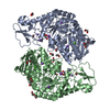
| Title | The structural and biochemical characterization of acyl-coa hydrolase mutant Asn28Ala from Staphylococcus aureus in complex with COENZYME A | ||||||
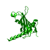
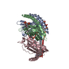
 Components Components | Acyl CoA Hydrolase | ||||||
 Keywords Keywords | HYDROLASE / Acyl CoA thioesterase / Staphylococcus aureus / Coenzyme A / Hotdog thioesterase | ||||||
| Function / homology |  Function and homology information Function and homology informationlong-chain fatty acyl-CoA hydrolase activity / acyl-CoA metabolic process / fatty acid catabolic process / cytosol Similarity search - Function | ||||||
| Biological species |   Staphylococcus aureus (bacteria) Staphylococcus aureus (bacteria) | ||||||
| Method |  X-RAY DIFFRACTION / SYNCHROTRON / MOLECULAR REPLACEMENT / Resolution: 2.4 Å X-RAY DIFFRACTION / SYNCHROTRON / MOLECULAR REPLACEMENT / Resolution: 2.4 Å | ||||||
 Authors Authors | Khandokar, Y.B. / Srivastava, P.S. / Forwood, J.K. | ||||||
 Citation Citation | Journal: To Be Published Title: The structural and biochemical characterization of acyl-coa hydrolase from Staphylococcus aureus Authors: Khandokar, Y.B. / Srivastava, P.S. / Forwood, J.K. | ||||||
| History |
|
- Structure visualization
| Structure viewer | Molecule: MolmilJmol/JSmol |
|---|
- Downloads & links
-Download
| PDBx/mmCIF format | 5egj.cif.gz | 50.3 KB | Display | PDBx/mmCIF format |
|---|---|---|---|---|
| PDB format | pdb5egj.ent.gz | 33.7 KB | Display | PDB format |
| PDBx/mmJSON format | 5egj.json.gz | Tree view | PDBx/mmJSON format | |
| Others |  Other downloads Other downloads |
-Validation report
| Arichive directory | https://data.pdbj.org/pub/pdb/validation_reports/eg/5egjftp://data.pdbj.org/pub/pdb/validation_reports/eg/5egj | HTTPS FTP |
|---|
-Related structure data
| Related structure data |  5egkC  5eglC  5hwfC 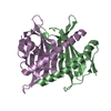 4ncpS C: citing same article ( S: Starting model for refinement |
|---|---|
| Similar structure data |

-Links
 PDBj
PDBj


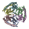
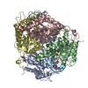
- Assembly
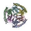
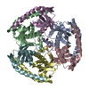
| Deposited unit | 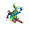
| ||||||||
|---|---|---|---|---|---|---|---|---|---|
| 1 | x 6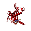
| ||||||||
| Unit cell |
| ||||||||
| Components on special symmetry positions |
|
-Components
| #1: Protein | Mass: 20221.857 Da / Num. of mol.: 1 Source method: isolated from a genetically manipulated source Source: (gene. exp.) Staphylococcus aureus (strain Mu50 / ATCC 700699) (bacteria)Strain: Mu50 / ATCC 700699 / Gene: SAV1878 / Plasmid: pMCSG21 / Production host: |
|---|---|
| #2: Chemical | ChemComp-COA /   Mass: 767.534 Da / Num. of mol.: 1 / Source method: obtained synthetically / Formula: C21H36N7O16P3S Mass: 767.534 Da / Num. of mol.: 1 / Source method: obtained synthetically / Formula: C21H36N7O16P3S |
| #3: Water | ChemComp-HOH /  Mass: 18.015 Da / Num. of mol.: 7 / Source method: isolated from a natural source / Formula: H2O Mass: 18.015 Da / Num. of mol.: 7 / Source method: isolated from a natural source / Formula: H2O |
-Experimental details
-Experiment
| Experiment | Method: X-RAY DIFFRACTION |
|---|
- Sample preparation
| Crystal | Density Matthews: 3.29 Å3/Da / Density % sol: 62 % |
|---|---|
| Crystal grow | Temperature: 296 K / Method: vapor diffusion, hanging drop / pH: 7.5 Details: 0.1M Sodium HEPES pH7.5, 0.8 M Sodium phosphate monobasic |
-Data collection
| Diffraction | Mean temperature: 100 K |
|---|---|
| Diffraction source | Source: SYNCHROTRON / Site: Australian Synchrotron  / Beamline: MX2 / Wavelength: 0.9537 Å / Beamline: MX2 / Wavelength: 0.9537 Å |
| Detector | Type: ADSC QUANTUM 315r / Detector: CCD / Date: Mar 31, 2015 |
| Radiation | Monochromator: SILICON DOUBLE CRYSTAL / Protocol: SINGLE WAVELENGTH / Monochromatic (M) / Laue (L): M / Scattering type: x-ray |
| Radiation wavelength | Wavelength: 0.9537 Å / Relative weight: 1 |
| Reflection | Resolution: 2.4→33.44 Å / Num. obs: 10919 / % possible obs: 99.93 % / Redundancy: 2 % / Rmerge(I) obs: 0.02896 / Net I/σ(I): 8.73 |
| Reflection shell | Resolution: 2.4→2.486 Å / Redundancy: 2 % / Rmerge(I) obs: 0.09755 / Mean I/σ(I) obs: 3.61 / % possible all: 100 |
- Processing
| Software |
| |||||||||||||||||||||||||||||||||||
|---|---|---|---|---|---|---|---|---|---|---|---|---|---|---|---|---|---|---|---|---|---|---|---|---|---|---|---|---|---|---|---|---|---|---|---|---|
| Refinement | Method to determine structure: MOLECULAR REPLACEMENT Starting model: 4NCP Resolution: 2.4→33.44 Å / SU ML: 0.27 / Cross valid method: FREE R-VALUE / σ(F): 1.34 / Phase error: 25.46 / Stereochemistry target values: ML
| |||||||||||||||||||||||||||||||||||
| Solvent computation | Shrinkage radii: 0.9 Å / VDW probe radii: 1.11 Å / Solvent model: FLAT BULK SOLVENT MODEL | |||||||||||||||||||||||||||||||||||
| Displacement parameters | Biso mean: 34.6 Å2 | |||||||||||||||||||||||||||||||||||
| Refinement step | Cycle: LAST / Resolution: 2.4→33.44 Å
| |||||||||||||||||||||||||||||||||||
| Refine LS restraints |
| |||||||||||||||||||||||||||||||||||
| LS refinement shell |
|
