 Movie
Movie Controller

[English] 日本語
 Yorodumi
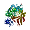
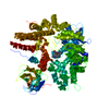
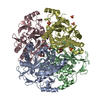
- PDB-1fiu: TETRAMERIC RESTRICTION ENDONUCLEASE NGOMIV IN COMPLEX WITH CLEAVED DNA -
+ Open data

- Basic information
| Entry | Database: PDB / ID: 1fiu | ||||||
|---|---|---|---|---|---|---|---|
| Title | TETRAMERIC RESTRICTION ENDONUCLEASE NGOMIV IN COMPLEX WITH CLEAVED DNA | ||||||
 Components Components |
| ||||||
 Keywords Keywords | hydrolase/DNA / PROTEIN-DNA COMPLEX / DOUBLE HELIX / RESTRICTION ENDONUCLEASE / RESTRICTION-MODIFICTION SYSTEMS / HYDROLASE / PHOSPHODIESTERASE / METAL ION / COMPLEX (ENDONUCLEASE-DNA) / hydrolase-DNA COMPLEX | ||||||
| Function / homology |  Function and homology information Function and homology informationtype II site-specific deoxyribonuclease / type II site-specific deoxyribonuclease activity / DNA restriction-modification system / metal ion binding Similarity search - Function | ||||||
| Biological species |  Neisseria gonorrhoeae (bacteria) Neisseria gonorrhoeae (bacteria) | ||||||
| Method |  X-RAY DIFFRACTION / SYNCHROTRON / Resolution: 1.6 Å X-RAY DIFFRACTION / SYNCHROTRON / Resolution: 1.6 Å | ||||||
 Authors Authors | Deibert, M. / Grazulis, S. / Sasnauskas, G. / Siksnys, V. / Huber, R. | ||||||
 Citation Citation | Journal: Nat.Struct.Biol. / Year: 2000 Title: Structure of the tetrameric restriction endonuclease NgoMIV in complex with cleaved DNA. Authors: Deibert, M. / Grazulis, S. / Sasnauskas, G. / Siksnys, V. / Huber, R. | ||||||
| History |
|
- Structure visualization
| Structure viewer | Molecule: MolmilJmol/JSmol |
|---|
- Downloads & links
-Download
| PDBx/mmCIF format | 1fiu.cif.gz | 289.1 KB | Display | PDBx/mmCIF format |
|---|---|---|---|---|
| PDB format | pdb1fiu.ent.gz | 227.1 KB | Display | PDB format |
| PDBx/mmJSON format | 1fiu.json.gz | Tree view | PDBx/mmJSON format | |
| Others |  Other downloads Other downloads |
-Validation report
| Arichive directory | https://data.pdbj.org/pub/pdb/validation_reports/fi/1fiuftp://data.pdbj.org/pub/pdb/validation_reports/fi/1fiu | HTTPS FTP |
|---|
-Related structure data
| Similar structure data |
|---|


-Links
 PDBj
PDBj


- Assembly
| Deposited unit | 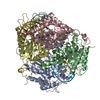
| ||||||||||
|---|---|---|---|---|---|---|---|---|---|---|---|
| 1 |
| ||||||||||
| Unit cell |
| ||||||||||
| Details | The biological assembly is a tetramer |
-Components
-DNA chain , 2 types, 8 molecules EFGHIJKL
| #1: DNA chain | Mass: 1206.829 Da / Num. of mol.: 4 / Source method: obtained synthetically #2: DNA chain | Mass: 2099.387 Da / Num. of mol.: 4 / Source method: obtained synthetically |
|---|
-Protein , 1 types, 4 molecules ABCD
| #3: Protein | Mass: 31810.902 Da / Num. of mol.: 4 Source method: isolated from a genetically manipulated source Source: (gene. exp.) Neisseria gonorrhoeae (bacteria) / Plasmid: PET15B / Production host: References: UniProt: P31032, type II site-specific deoxyribonuclease |
|---|
-Non-polymers , 3 types, 1291 molecules 


| #4: Chemical | ChemComp-MG /  Mass: 24.305 Da / Num. of mol.: 8 / Source method: obtained synthetically / Formula: Mg Mass: 24.305 Da / Num. of mol.: 8 / Source method: obtained synthetically / Formula: Mg#5: Chemical | ChemComp-ACY /  Mass: 60.052 Da / Num. of mol.: 4 / Source method: obtained synthetically / Formula: C2H4O2 Mass: 60.052 Da / Num. of mol.: 4 / Source method: obtained synthetically / Formula: C2H4O2#6: Water | ChemComp-HOH / | Mass: 18.015 Da / Num. of mol.: 1279 / Source method: isolated from a natural source / Formula: H2O |
|---|
-Experimental details
-Experiment
| Experiment | Method: X-RAY DIFFRACTION / Number of used crystals: 1 |
|---|
- Sample preparation
| Crystal | Density Matthews: 2.18 Å3/Da / Density % sol: 43.62 % | ||||||||||||||||||||||||||||||||||||||||||||||||||||||||
|---|---|---|---|---|---|---|---|---|---|---|---|---|---|---|---|---|---|---|---|---|---|---|---|---|---|---|---|---|---|---|---|---|---|---|---|---|---|---|---|---|---|---|---|---|---|---|---|---|---|---|---|---|---|---|---|---|---|
| Crystal grow | Temperature: 291 K / Method: vapor diffusion, sitting drop / pH: 6.5 Details: MES 100mM, MgCl2 200mM, MPD 25%, pH 6.5, VAPOR DIFFUSION, SITTING DROP, temperature 291K | ||||||||||||||||||||||||||||||||||||||||||||||||||||||||
| Components of the solutions |
| ||||||||||||||||||||||||||||||||||||||||||||||||||||||||
| Crystal grow | *PLUS pH: 7.5 | ||||||||||||||||||||||||||||||||||||||||||||||||||||||||
| Components of the solutions | *PLUS
|
-Data collection
| Diffraction | Mean temperature: 90 K |
|---|---|
| Diffraction source | Source: SYNCHROTRON / Site: MPG/DESY, HAMBURG  / Beamline: BW6 / Wavelength: 1.0697 / Beamline: BW6 / Wavelength: 1.0697 |
| Detector | Type: MARRESEARCH / Detector: IMAGE PLATE / Date: Jul 21, 1998 |
| Radiation | Protocol: SINGLE WAVELENGTH / Monochromatic (M) / Laue (L): M / Scattering type: x-ray |
| Radiation wavelength | Wavelength: 1.0697 Å / Relative weight: 1 |
| Reflection | Resolution: 1.6→17.26 Å / Num. all: 162845 / Num. obs: 160528 / % possible obs: 98.6 % / Observed criterion σ(I): 1 / Redundancy: 15.7 % / Rmerge(I) obs: 0.047 |
| Reflection shell | Highest resolution: 1.6 Å / % possible all: 88.7 |
| Reflection | *PLUS Num. measured all: 2553379 |
| Reflection shell | *PLUS % possible obs: 88.7 % |
- Processing
| Software |
| |||||||||||||||||||||||||
|---|---|---|---|---|---|---|---|---|---|---|---|---|---|---|---|---|---|---|---|---|---|---|---|---|---|---|
| Refinement | Resolution: 1.6→17.26 Å / Cross valid method: THROUGHOUT / σ(F): 2 / Stereochemistry target values: Engh & Huber
| |||||||||||||||||||||||||
| Refinement step | Cycle: LAST / Resolution: 1.6→17.26 Å
| |||||||||||||||||||||||||
| Refine LS restraints |
| |||||||||||||||||||||||||
| Software | *PLUS Name: X-PLOR / Classification: refinement | |||||||||||||||||||||||||
| Refinement | *PLUS Highest resolution: 1.6 Å / σ(F): 2 / % reflection Rfree: 10 % / Rfactor obs: 0.174 | |||||||||||||||||||||||||
| Solvent computation | *PLUS | |||||||||||||||||||||||||
| Displacement parameters | *PLUS | |||||||||||||||||||||||||
| Refine LS restraints | *PLUS
|
