determination of heart left/right asymmetry / blood vessel endothelial cell fate specification / positive regulation of ERBB signaling pathway / club cell differentiation / arterial endothelial cell fate commitment / blood vessel lumenization / regulation of generation of precursor metabolites and energy / regulation of timing of cell differentiation / positive regulation of ephrin receptor signaling pathway / pulmonary valve development ...determination of heart left/right asymmetry / blood vessel endothelial cell fate specification / positive regulation of ERBB signaling pathway / club cell differentiation / arterial endothelial cell fate commitment / blood vessel lumenization / regulation of generation of precursor metabolites and energy / regulation of timing of cell differentiation / positive regulation of ephrin receptor signaling pathway / pulmonary valve development / dorsal aorta morphogenesis / sebaceous gland development / positive regulation of cell proliferation involved in heart morphogenesis / endocardium morphogenesis / MAML1-RBP-Jkappa- ICN1 complex / NOTCH1 Intracellular Domain Regulates Transcription / RUNX3 regulates NOTCH signaling / secondary heart field specification / auditory receptor cell fate commitment / positive regulation of transcription of Notch receptor target / aortic valve development / Notch-HLH transcription pathway / heart induction / epithelial to mesenchymal transition involved in endocardial cushion formation / epidermal cell fate specification / pituitary gland development / cardiac left ventricle morphogenesis / endocardium development / regulation of cell adhesion involved in heart morphogenesis / atrioventricular canal development / hair follicle maturation / cardiac muscle cell myoblast differentiation / myeloid dendritic cell differentiation / ventricular trabecula myocardium morphogenesis / positive regulation of BMP signaling pathway / regulation of epithelial cell proliferation / inflammatory response to antigenic stimulus / nuclear export / negative regulation of ossification / negative regulation of cold-induced thermogenesis / artery morphogenesis / labyrinthine layer blood vessel development / ventricular septum morphogenesis / heart looping / outflow tract morphogenesis / humoral immune response / hemopoiesis / negative regulation of cell differentiation / somatic stem cell population maintenance / epithelial to mesenchymal transition / blood vessel remodeling / negative regulation of Notch signaling pathway / cell fate commitment / somitogenesis / negative regulation of stem cell proliferation / keratinocyte differentiation / positive regulation of cardiac muscle cell proliferation / neurogenesis / Notch signaling pathway / transcription repressor complex / tubulin binding / B cell differentiation / epithelial cell proliferation / positive regulation of epithelial cell proliferation / stem cell proliferation / neuron differentiation / positive regulation of canonical Wnt signaling pathway / regulation of gene expression / heart development / DNA-binding transcription activator activity, RNA polymerase II-specific / angiogenesis / transcription regulator complex / sequence-specific DNA binding / RNA polymerase II-specific DNA-binding transcription factor binding / cell differentiation / cell population proliferation / defense response to bacterium / RNA polymerase II cis-regulatory region sequence-specific DNA binding / negative regulation of DNA-templated transcription / centrosome / chromatin binding / positive regulation of gene expression / regulation of transcription by RNA polymerase II / positive regulation of DNA-templated transcription / nucleolus / negative regulation of transcription by RNA polymerase II / positive regulation of transcription by RNA polymerase II / DNA binding / nucleoplasm / nucleus / cytoplasm Similarity search - Function
RBPJ-interacting and tubulin-associated protein 1 / RBPJ-interacting and tubulin associated protein / LAG1, DNA binding domain / Beta-trefoil DNA-binding domain / RBP-J/Cbf11/Cbf12, DNA binding / Beta-trefoil domain superfamily / RBP-J/Cbf11, DNA binding domain superfamily / RBP-Jkappa, IPT domain / Suppressor of hairless-like / Beta-trefoil DNA-binding domain ...RBPJ-interacting and tubulin-associated protein 1 / RBPJ-interacting and tubulin associated protein / LAG1, DNA binding domain / Beta-trefoil DNA-binding domain / RBP-J/Cbf11/Cbf12, DNA binding / Beta-trefoil domain superfamily / RBP-J/Cbf11, DNA binding domain superfamily / RBP-Jkappa, IPT domain / Suppressor of hairless-like / Beta-trefoil DNA-binding domain / LAG1, DNA binding / TIG domain / LAG1, DNA binding / Beta-trefoil DNA-binding domain / p53-like transcription factor, DNA-binding / Trefoil (Acidic Fibroblast Growth Factor, subunit A) - #50 / Trefoil (Acidic Fibroblast Growth Factor, subunit A) / Trefoil / Immunoglobulin E-set / Immunoglobulin-like fold / Immunoglobulins / Immunoglobulin-like / Sandwich / Mainly Beta Similarity search - Domain/homology
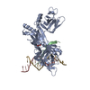
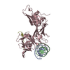
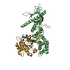
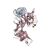
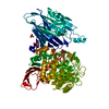
1,4-BUTANEDIOL / DNA / DNA (> 10) / Recombining binding protein suppressor of hairless / RBPJ-interacting and tubulin-associated protein 1 Similarity search - Component
B: DNA (5'-D(*AP*AP*TP*CP*TP*TP*TP*CP*CP*CP*AP*CP*AP*GP*T)-3') C: Recombining binding protein suppressor of hairless R: hRITA A: DNA (5'-D(*TP*TP*AP*CP*TP*GP*TP*GP*GP*GP*AP*AP*AP*GP*A)-3') hetero molecules
In the structure databanks used in Yorodumi, some data are registered as the other names, "COVID-19 virus" and "2019-nCoV". Here are the details of the virus and the list of structure data.
Jan 31, 2019. EMDB accession codes are about to change! (news from PDBe EMDB page)
EMDB accession codes are about to change! (news from PDBe EMDB page)
The allocation of 4 digits for EMDB accession codes will soon come to an end. Whilst these codes will remain in use, new EMDB accession codes will include an additional digit and will expand incrementally as the available range of codes is exhausted. The current 4-digit format prefixed with “EMD-” (i.e. EMD-XXXX) will advance to a 5-digit format (i.e. EMD-XXXXX), and so on. It is currently estimated that the 4-digit codes will be depleted around Spring 2019, at which point the 5-digit format will come into force.
The EM Navigator/Yorodumi systems omit the EMD- prefix.
Related info.:Q: What is EMD? / ID/Accession-code notation in Yorodumi/EM Navigator
Yorodumi is a browser for structure data from EMDB, PDB, SASBDB, etc.
This page is also the successor to EM Navigator detail page, and also detail information page/front-end page for Omokage search.
The word "yorodu" (or yorozu) is an old Japanese word meaning "ten thousand". "mi" (miru) is to see.
Related info.:EMDB / PDB / SASBDB / Comparison of 3 databanks / Yorodumi Search / Aug 31, 2016. New EM Navigator & Yorodumi / Yorodumi Papers / Jmol/JSmol / Function and homology information / Changes in new EM Navigator and Yorodumi
 Movie
Movie Controller


 Open data
 Basic information
 Components
 Keywords
 Function and homology information
 X-RAY DIFFRACTION /
  Authors
 United States, 2items
  Citation
 Structure visualization
 Downloads & links
 Other downloads
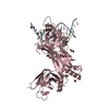


 PDBj
PDBj


 Assembly
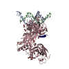


 Mass: 62.068 Da / Num. of mol.: 19 / Source method: obtained synthetically / Formula: C2H6O2
 Mass: 90.121 Da / Num. of mol.: 7 / Source method: obtained synthetically / Formula: C4H10O2
 Sample preparation
 Processing