- PDB-5eg1: Antibacterial peptide ABC transporter McjD with a resolved lipid -
+
Open data
ID or keywords:
Loading...
-
Basic information
Entry
Database: PDB / ID: 5eg1
Title
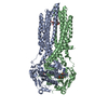
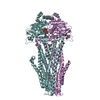
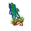
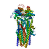
Antibacterial peptide ABC transporter McjD with a resolved lipid
Components
Microcin-J25 export ATP-binding/permease protein McjD
Keywords
TRANSPORT PROTEIN / membrane protein / ABC transporter / lipid
Function / homology
Function and homology information
bacteriocin transport / bacteriocin immunity / ABC-type oligopeptide transporter activity / protein transport / ATP hydrolysis activity / ATP binding / plasma membrane Similarity search - Function
Type 1 protein exporter / ABC transporter transmembrane region / ABC transporter type 1, transmembrane domain / ABC transporter integral membrane type-1 fused domain profile. / ABC transporter type 1, transmembrane domain superfamily / ABC transporter-like, conserved site / ABC transporters family signature. / ABC transporter / ABC transporter-like, ATP-binding domain / ATP-binding cassette, ABC transporter-type domain profile. ...Type 1 protein exporter / ABC transporter transmembrane region / ABC transporter type 1, transmembrane domain / ABC transporter integral membrane type-1 fused domain profile. / ABC transporter type 1, transmembrane domain superfamily / ABC transporter-like, conserved site / ABC transporters family signature. / ABC transporter / ABC transporter-like, ATP-binding domain / ATP-binding cassette, ABC transporter-type domain profile. / P-loop containing nucleotide triphosphate hydrolases / ATPases associated with a variety of cellular activities / AAA+ ATPase domain / Rossmann fold / P-loop containing nucleoside triphosphate hydrolase / 3-Layer(aba) Sandwich / Alpha Beta Similarity search - Domain/homology
Resolution: 3.42→67.61 Å / Cor.coef. Fo:Fc: 0.7807 / Cor.coef. Fo:Fc free: 0.809 / Cross valid method: THROUGHOUT / σ(F): 0 / SU Rfree Blow DPI: 0.513 Details: Due to low resolution of the data, the ligand head group could either a glycerol or ethanolamine moiety. We assigned it as glycerol since it was supported by our biochemical data. Both head ...Details: Due to low resolution of the data, the ligand head group could either a glycerol or ethanolamine moiety. We assigned it as glycerol since it was supported by our biochemical data. Both head groups can be refined without the presence of any negative Fo-Fc density
Rfactor
Num. reflection
% reflection
Selection details
Rfree
0.2718
1458
5.02 %
RANDOM
Rwork
0.2535
-
-
-
obs
0.2544
29028
98.7 %
-
Displacement parameters
Biso mean: 126.14 Å2
Baniso -1
Baniso -2
Baniso -3
1-
-62.3634 Å2
0 Å2
0 Å2
2-
-
-17.7063 Å2
0 Å2
3-
-
-
80.0697 Å2
Refine analyze
Luzzati coordinate error obs: 1.298 Å
Refinement step
Cycle: 1 / Resolution: 3.42→67.61 Å
Protein
Nucleic acid
Ligand
Solvent
Total
Num. atoms
9107
0
115
0
9222
Refine LS restraints
Refine-ID
Type
Dev ideal
Number
Restraint function
Weight
X-RAY DIFFRACTION
t_bond_d
0.01
9378
HARMONIC
2
X-RAY DIFFRACTION
t_angle_deg
1.07
12705
HARMONIC
2
X-RAY DIFFRACTION
t_dihedral_angle_d
4452
SINUSOIDAL
2
X-RAY DIFFRACTION
t_incorr_chiral_ct
X-RAY DIFFRACTION
t_pseud_angle
X-RAY DIFFRACTION
t_trig_c_planes
216
HARMONIC
2
X-RAY DIFFRACTION
t_gen_planes
1360
HARMONIC
5
X-RAY DIFFRACTION
t_it
9378
HARMONIC
20
X-RAY DIFFRACTION
t_nbd
2
SEMIHARMONIC
5
X-RAY DIFFRACTION
t_omega_torsion
2.42
X-RAY DIFFRACTION
t_other_torsion
3.62
X-RAY DIFFRACTION
t_improper_torsion
X-RAY DIFFRACTION
t_chiral_improper_torsion
1281
SEMIHARMONIC
5
X-RAY DIFFRACTION
t_sum_occupancies
X-RAY DIFFRACTION
t_utility_distance
X-RAY DIFFRACTION
t_utility_angle
X-RAY DIFFRACTION
t_utility_torsion
X-RAY DIFFRACTION
t_ideal_dist_contact
11154
SEMIHARMONIC
4
LS refinement shell
Resolution: 3.42→3.54 Å / Total num. of bins used: 15
Rfactor
Num. reflection
% reflection
Rfree
0.3042
145
5.19 %
Rwork
0.2998
2649
-
all
0.3
2794
-
obs
-
-
98.7 %
+
About Yorodumi
-
News
-
Feb 9, 2022. New format data for meta-information of EMDB entries
New format data for meta-information of EMDB entries
Version 3 of the EMDB header file is now the official format.
The previous official version 1.9 will be removed from the archive.
In the structure databanks used in Yorodumi, some data are registered as the other names, "COVID-19 virus" and "2019-nCoV". Here are the details of the virus and the list of structure data.
Jan 31, 2019. EMDB accession codes are about to change! (news from PDBe EMDB page)
EMDB accession codes are about to change! (news from PDBe EMDB page)
The allocation of 4 digits for EMDB accession codes will soon come to an end. Whilst these codes will remain in use, new EMDB accession codes will include an additional digit and will expand incrementally as the available range of codes is exhausted. The current 4-digit format prefixed with “EMD-” (i.e. EMD-XXXX) will advance to a 5-digit format (i.e. EMD-XXXXX), and so on. It is currently estimated that the 4-digit codes will be depleted around Spring 2019, at which point the 5-digit format will come into force.
The EM Navigator/Yorodumi systems omit the EMD- prefix.
Related info.:Q: What is EMD? / ID/Accession-code notation in Yorodumi/EM Navigator
Yorodumi is a browser for structure data from EMDB, PDB, SASBDB, etc.
This page is also the successor to EM Navigator detail page, and also detail information page/front-end page for Omokage search.
The word "yorodu" (or yorozu) is an old Japanese word meaning "ten thousand". "mi" (miru) is to see.
Related info.:EMDB / PDB / SASBDB / Comparison of 3 databanks / Yorodumi Search / Aug 31, 2016. New EM Navigator & Yorodumi / Yorodumi Papers / Jmol/JSmol / Function and homology information / Changes in new EM Navigator and Yorodumi
 Movie
Movie Controller


 Open data
 Basic information
 Components
 Keywords
 Function and homology information
 X-RAY DIFFRACTION /
  Authors
 United Kingdom, 1items
  Citation
 Structure visualization
 Downloads & links
 Other downloads


 PDBj
PDBj


 Assembly
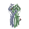

 Mass: 506.196 Da / Num. of mol.: 2 / Source method: obtained synthetically / Formula: C10H17N6O12P3 / Comment: AMP-PNP, energy-carrying molecule analogue*YM
 Mass: 24.305 Da / Num. of mol.: 2 / Source method: obtained synthetically / Formula: Mg
 Mass: 746.991 Da / Num. of mol.: 1 / Source method: obtained synthetically / Formula: C40H75O10P / Comment: phospholipid*YM
 Sample preparation
 Processing