- PDB-4pl0: Crystal structure of the antibacterial peptide ABC transporter Mc... -
+
Open data
ID or keywords:
Loading...
-
Basic information
Entry
Database: PDB / ID: 4pl0
Title
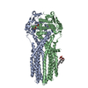
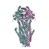
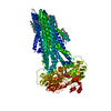
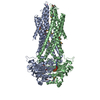
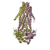
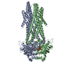
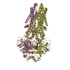
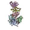
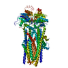
Crystal structure of the antibacterial peptide ABC transporter McjD in an outward occluded state
Components
Microcin-J25 export ATP-binding/permease protein McjD
Keywords
TRANSPORT PROTEIN / ABC transporter / membrane protein / occluded
Function / homology
Function and homology information
bacteriocin transport / bacteriocin immunity / ABC-type oligopeptide transporter activity / protein transport / ATP hydrolysis activity / ATP binding / plasma membrane Similarity search - Function
Type 1 protein exporter / ABC transporter transmembrane region / ABC transporter type 1, transmembrane domain / ABC transporter integral membrane type-1 fused domain profile. / ABC transporter type 1, transmembrane domain superfamily / ABC transporter-like, conserved site / ABC transporters family signature. / ABC transporter / ABC transporter-like, ATP-binding domain / ATP-binding cassette, ABC transporter-type domain profile. ...Type 1 protein exporter / ABC transporter transmembrane region / ABC transporter type 1, transmembrane domain / ABC transporter integral membrane type-1 fused domain profile. / ABC transporter type 1, transmembrane domain superfamily / ABC transporter-like, conserved site / ABC transporters family signature. / ABC transporter / ABC transporter-like, ATP-binding domain / ATP-binding cassette, ABC transporter-type domain profile. / P-loop containing nucleotide triphosphate hydrolases / ATPases associated with a variety of cellular activities / AAA+ ATPase domain / Rossmann fold / P-loop containing nucleoside triphosphate hydrolase / 3-Layer(aba) Sandwich / Alpha Beta Similarity search - Domain/homology
In the structure databanks used in Yorodumi, some data are registered as the other names, "COVID-19 virus" and "2019-nCoV". Here are the details of the virus and the list of structure data.
Jan 31, 2019. EMDB accession codes are about to change! (news from PDBe EMDB page)
EMDB accession codes are about to change! (news from PDBe EMDB page)
The allocation of 4 digits for EMDB accession codes will soon come to an end. Whilst these codes will remain in use, new EMDB accession codes will include an additional digit and will expand incrementally as the available range of codes is exhausted. The current 4-digit format prefixed with “EMD-” (i.e. EMD-XXXX) will advance to a 5-digit format (i.e. EMD-XXXXX), and so on. It is currently estimated that the 4-digit codes will be depleted around Spring 2019, at which point the 5-digit format will come into force.
The EM Navigator/Yorodumi systems omit the EMD- prefix.
Related info.:Q: What is EMD? / ID/Accession-code notation in Yorodumi/EM Navigator
Yorodumi is a browser for structure data from EMDB, PDB, SASBDB, etc.
This page is also the successor to EM Navigator detail page, and also detail information page/front-end page for Omokage search.
The word "yorodu" (or yorozu) is an old Japanese word meaning "ten thousand". "mi" (miru) is to see.
Related info.:EMDB / PDB / SASBDB / Comparison of 3 databanks / Yorodumi Search / Aug 31, 2016. New EM Navigator & Yorodumi / Yorodumi Papers / Jmol/JSmol / Function and homology information / Changes in new EM Navigator and Yorodumi
 Movie
Movie Controller

 Yorodumi
 Open data
 Basic information
 Components
 Keywords
 Function and homology information
 X-RAY DIFFRACTION /
  Authors
 Citation
 Structure visualization
 Downloads & links
 Other downloads
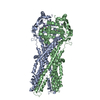

 PDBj
PDBj


 Assembly
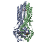

 Mass: 506.196 Da / Num. of mol.: 2 / Source method: obtained synthetically / Formula: C10H17N6O12P3 / Comment: AMP-PNP, energy-carrying molecule analogue*YM
 Mass: 24.305 Da / Num. of mol.: 2 / Source method: obtained synthetically / Formula: Mg
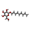
 Type: D-saccharide / Mass: 306.395 Da / Num. of mol.: 2 / Source method: obtained synthetically / Formula: C15H30O6 / Comment: detergent*YM
 Sample preparation
 / Beamline: I02 / Wavelength: 0.979 Å
 Processing