 Movie
Movie Controller


+ Open data

- Basic information
| Entry | Database: PDB / ID: 5e5t | ||||||
|---|---|---|---|---|---|---|---|
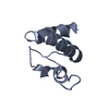
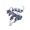
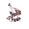
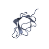
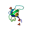
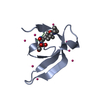
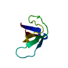
| Title | Quasi-racemic snakin-1 in P1 after radiation damage | ||||||
 Components Components |
| ||||||
 Keywords Keywords | ANTIMICROBIAL PROTEIN / GASA/snakin / cysteine-rich antimicrobial peptide | ||||||
| Function / homology | Gibberellin regulated protein / Gibberellin regulated protein / defense response / extracellular region / FORMIC ACID / Snakin-1 / Snakin-1 Function and homology information Function and homology information | ||||||
| Biological species |  | ||||||
| Method |  X-RAY DIFFRACTION / SYNCHROTRON / MIR / Resolution: 1.572 Å X-RAY DIFFRACTION / SYNCHROTRON / MIR / Resolution: 1.572 Å | ||||||
 Authors Authors | Yeung, H. / Squire, C.J. / Yosaatmadja, Y. / Panjikar, S. / Baker, E.N. / Harris, P.W.R. / Brimble, M.A. | ||||||
| Funding support |  New Zealand, 1items New Zealand, 1items
| ||||||
 Citation Citation | Journal: Angew.Chem.Int.Ed.Engl. / Year: 2016 Title: Radiation Damage and Racemic Protein Crystallography Reveal the Unique Structure of the GASA/Snakin Protein Superfamily. Authors: Yeung, H. / Squire, C.J. / Yosaatmadja, Y. / Panjikar, S. / Lopez, G. / Molina, A. / Baker, E.N. / Harris, P.W. / Brimble, M.A. | ||||||
| History |
|
- Structure visualization
| Structure viewer | Molecule: MolmilJmol/JSmol |
|---|
- Downloads & links
-Download
| PDBx/mmCIF format | 5e5t.cif.gz | 71.7 KB | Display | PDBx/mmCIF format |
|---|---|---|---|---|
| PDB format | pdb5e5t.ent.gz | 57.1 KB | Display | PDB format |
| PDBx/mmJSON format | 5e5t.json.gz | Tree view | PDBx/mmJSON format | |
| Others |  Other downloads Other downloads |
-Validation report
| Summary document | 5e5t_validation.pdf.gz | 458.4 KB | Display | wwPDB validaton report |
|---|---|---|---|---|
| Full document | 5e5t_full_validation.pdf.gz | 458.5 KB | Display | |
| Data in XML | 5e5t_validation.xml.gz | 13.3 KB | Display | |
| Data in CIF | 5e5t_validation.cif.gz | 18.9 KB | Display | |
| Arichive directory | https://data.pdbj.org/pub/pdb/validation_reports/e5/5e5tftp://data.pdbj.org/pub/pdb/validation_reports/e5/5e5t | HTTPS FTP |
-Related structure data


-Links
 PDBj
PDBj- Assembly
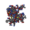
| Deposited unit | 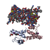
| ||||||||
|---|---|---|---|---|---|---|---|---|---|
| 1 | 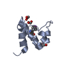
| ||||||||
| 2 | 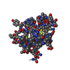
| ||||||||
| 3 | 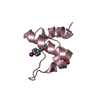
| ||||||||
| 4 | 
| ||||||||
| Unit cell |
|
-Components
| #1: Protein | Mass: 7059.083 Da / Num. of mol.: 2 / Mutation: Y25(PHI) / Source method: obtained synthetically Details: Potato L- snakin-1 containing single substitution of p-iodophenylalanine for 25Tyr Source: (synth.) #2: Protein | Mass: 6938.109 Da / Num. of mol.: 2 / Source method: obtained synthetically / Details: D- enantiomer of potato snakin-1 / Source: (synth.) #3: Chemical | ChemComp-FMT /   Mass: 46.025 Da / Num. of mol.: 4 / Source method: obtained synthetically / Formula: CH2O2 Mass: 46.025 Da / Num. of mol.: 4 / Source method: obtained synthetically / Formula: CH2O2#4: Chemical |   Mass: 62.068 Da / Num. of mol.: 3 / Source method: obtained synthetically / Formula: C2H6O2 Mass: 62.068 Da / Num. of mol.: 3 / Source method: obtained synthetically / Formula: C2H6O2#5: Water | ChemComp-HOH / |  Mass: 18.015 Da / Num. of mol.: 247 / Source method: isolated from a natural source / Formula: H2O Mass: 18.015 Da / Num. of mol.: 247 / Source method: isolated from a natural source / Formula: H2OHas protein modification | Y | |
|---|
-Experimental details
-Experiment
| Experiment | Method: X-RAY DIFFRACTION / Number of used crystals: 1 |
|---|
- Sample preparation
| Crystal | Density Matthews: 2.05 Å3/Da / Density % sol: 40.08 % |
|---|---|
| Crystal grow | Temperature: 291 K / Method: vapor diffusion, hanging drop / Details: 3.8 M sodium formate |
-Data collection
| Diffraction | Mean temperature: 100 K |
|---|---|
| Diffraction source | Source: SYNCHROTRON / Site: Australian Synchrotron  / Beamline: MX2 / Wavelength: 0.9919 Å / Beamline: MX2 / Wavelength: 0.9919 Å |
| Detector | Type: ADSC QUANTUM 315r / Detector: CCD / Date: Apr 24, 2015 |
| Radiation | Protocol: SINGLE WAVELENGTH / Monochromatic (M) / Laue (L): M / Scattering type: x-ray |
| Radiation wavelength | Wavelength: 0.9919 Å / Relative weight: 1 |
| Reflection | Resolution: 1.57→36.49 Å / Num. obs: 29896 / % possible obs: 96.5 % / Observed criterion σ(I): 0 / Redundancy: 2.4 % / Rmerge(I) obs: 0.068 / Net I/σ(I): 8.4 |
| Reflection shell | Resolution: 1.57→1.6 Å / Redundancy: 2.4 % / Rmerge(I) obs: 0.513 / Mean I/σ(I) obs: 1.7 / % possible all: 93.2 |
- Processing
| Software |
| ||||||||||||||||||||||||||||||||||||||||||||||||||||||||||||||||||||||||||||||||||||
|---|---|---|---|---|---|---|---|---|---|---|---|---|---|---|---|---|---|---|---|---|---|---|---|---|---|---|---|---|---|---|---|---|---|---|---|---|---|---|---|---|---|---|---|---|---|---|---|---|---|---|---|---|---|---|---|---|---|---|---|---|---|---|---|---|---|---|---|---|---|---|---|---|---|---|---|---|---|---|---|---|---|---|---|---|---|
| Refinement | Method to determine structure: MIR / Resolution: 1.572→36.488 Å / SU ML: 0.18 / Cross valid method: FREE R-VALUE / σ(F): 1.98 / Phase error: 27.76 / Stereochemistry target values: ML
| ||||||||||||||||||||||||||||||||||||||||||||||||||||||||||||||||||||||||||||||||||||
| Solvent computation | Shrinkage radii: 0.9 Å / VDW probe radii: 1.11 Å / Solvent model: FLAT BULK SOLVENT MODEL | ||||||||||||||||||||||||||||||||||||||||||||||||||||||||||||||||||||||||||||||||||||
| Refinement step | Cycle: LAST / Resolution: 1.572→36.488 Å
| ||||||||||||||||||||||||||||||||||||||||||||||||||||||||||||||||||||||||||||||||||||
| Refine LS restraints |
| ||||||||||||||||||||||||||||||||||||||||||||||||||||||||||||||||||||||||||||||||||||
| LS refinement shell |
|
