 Movie
Movie Controller

[English] 日本語
 Yorodumi
- PDB-5e1l: Structural and functional analysis of the E. coli FtsZ interactin... -
+ Open data

- Basic information
| Entry | Database: PDB / ID: 5e1l | ||||||
|---|---|---|---|---|---|---|---|
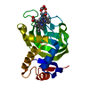
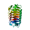
| Title | Structural and functional analysis of the E. coli FtsZ interacting protein, ZapC, reveals insight into molecular properties of a novel Z ring stabilizing protein | ||||||
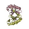
 Components Components | Cell division protein ZapC | ||||||
 Keywords Keywords | CELL CYCLE / Z ring / cell division | ||||||
| Function / homology |  Function and homology information Function and homology informationdivision septum assembly / FtsZ-dependent cytokinesis / cell division site / regulation of cell division / identical protein binding / cytoplasm Similarity search - Function | ||||||
| Biological species |  | ||||||
| Method |  X-RAY DIFFRACTION / SYNCHROTRON / SAD / MAD / Resolution: 2.15 Å X-RAY DIFFRACTION / SYNCHROTRON / SAD / MAD / Resolution: 2.15 Å | ||||||
 Authors Authors | Schumacher, M.A. / Huang, K.-H. / Tchorzewski, L. / Zeng, W. / Janakiraman, A. | ||||||
 Citation Citation | Journal: J.Biol.Chem. / Year: 2016 Title: Structural and Functional Analyses Reveal Insights into the Molecular Properties of the Escherichia coli Z Ring Stabilizing Protein, ZapC. Authors: Schumacher, M.A. / Zeng, W. / Huang, K.H. / Tchorzewski, L. / Janakiraman, A. | ||||||
| History |
|
- Structure visualization
| Structure viewer | Molecule: MolmilJmol/JSmol |
|---|
- Downloads & links
-Download
| PDBx/mmCIF format | 5e1l.cif.gz | 51 KB | Display | PDBx/mmCIF format |
|---|---|---|---|---|
| PDB format | pdb5e1l.ent.gz | 35.2 KB | Display | PDB format |
| PDBx/mmJSON format | 5e1l.json.gz | Tree view | PDBx/mmJSON format | |
| Others |  Other downloads Other downloads |
-Validation report
| Arichive directory | https://data.pdbj.org/pub/pdb/validation_reports/e1/5e1lftp://data.pdbj.org/pub/pdb/validation_reports/e1/5e1l | HTTPS FTP |
|---|
-Related structure data
| Similar structure data |
|---|


-Links
 PDBj
PDBj- Assembly
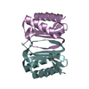
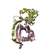
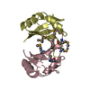
| Deposited unit | 
| ||||||||
|---|---|---|---|---|---|---|---|---|---|
| 1 |
| ||||||||
| Unit cell |
|
-Components
| #1: Protein | Mass: 22783.135 Da / Num. of mol.: 1 Source method: isolated from a genetically manipulated source Source: (gene. exp.) Gene: ycbW, zapC, AC789_1c09860, BN1008_2209, ECONIH1_05985, EL75_2796, EL79_2872, EL80_2810, HUS2011_1029, HW43_08455, IY32_15190, PD07_27680, PGD_02344, PU06_12140, PU21_19080, PU69_14370, RR31_ ...Gene: ycbW, zapC, AC789_1c09860, BN1008_2209, ECONIH1_05985, EL75_2796, EL79_2872, EL80_2810, HUS2011_1029, HW43_08455, IY32_15190, PD07_27680, PGD_02344, PU06_12140, PU21_19080, PU69_14370, RR31_05140, UN86_22105, UN92_26695, WQ64_03785, WQ65_11505, WQ66_20855, WQ69_14055, WQ70_10055, WQ71_17640, WQ72_12045, WQ73_04655, WQ74_18750, WQ77_23190, WQ79_17420, WQ84_15610, WQ86_15290, WQ88_01710, WQ92_17480, WQ95_04390, WQ99_00770, WR00_01300, WR01_18315, WR03_21275, WR05_24825, WR12_12790, WR13_00430, WR16_17045, WR17_22160, WR18_05905, WR19_22835, WR23_12280, WR24_08325, XB00_15425 Production host: |
|---|---|
| #2: Water | ChemComp-HOH /  Mass: 18.015 Da / Num. of mol.: 100 / Source method: isolated from a natural source / Formula: H2O Mass: 18.015 Da / Num. of mol.: 100 / Source method: isolated from a natural source / Formula: H2O |
-Experimental details
-Experiment
| Experiment | Method: X-RAY DIFFRACTION / Number of used crystals: 1 |
|---|
- Sample preparation
| Crystal | Density Matthews: 2.37 Å3/Da / Density % sol: 48.01 % |
|---|---|
| Crystal grow | Temperature: 298 K / Method: vapor diffusion, hanging drop / Details: 1.5 M sodium citrate, 0.1 M cacodylate pH 6.5 / PH range: 6.5 |
-Data collection
| Diffraction | Mean temperature: 100 K |
|---|---|
| Diffraction source | Source: SYNCHROTRON / Site: ALS  / Beamline: 8.3.1 / Wavelength: 1 Å / Beamline: 8.3.1 / Wavelength: 1 Å |
| Detector | Type: ADSC QUANTUM 315r / Detector: CCD / Date: Dec 21, 2013 |
| Radiation | Protocol: SINGLE WAVELENGTH / Monochromatic (M) / Laue (L): M / Scattering type: x-ray |
| Radiation wavelength | Wavelength: 1 Å / Relative weight: 1 |
| Reflection | Resolution: 2.15→60.5 Å / Num. obs: 9961 / % possible obs: 98 % / Observed criterion σ(F): 0 / Observed criterion σ(I): 0 / Redundancy: 3 % / Rsym value: 0.02 / Net I/σ(I): 11.5 |
| Reflection shell | Rmerge(I) obs: 0.234 / Mean I/σ(I) obs: 4.6 |
-Phasing
| Phasing | Method: MAD |
|---|
- Processing
| Software |
| ||||||||||||||||||||||||||||||||||||||||||||||||||||||||
|---|---|---|---|---|---|---|---|---|---|---|---|---|---|---|---|---|---|---|---|---|---|---|---|---|---|---|---|---|---|---|---|---|---|---|---|---|---|---|---|---|---|---|---|---|---|---|---|---|---|---|---|---|---|---|---|---|---|
| Refinement | Method to determine structure: SAD / Resolution: 2.15→37.643 Å / SU ML: 0.27 / Cross valid method: FREE R-VALUE / σ(F): 0 / Phase error: 24.54 / Stereochemistry target values: ML
| ||||||||||||||||||||||||||||||||||||||||||||||||||||||||
| Solvent computation | Shrinkage radii: 0.83 Å / VDW probe radii: 1.1 Å / Solvent model: FLAT BULK SOLVENT MODEL / Bsol: 40.377 Å2 / ksol: 0.344 e/Å3 | ||||||||||||||||||||||||||||||||||||||||||||||||||||||||
| Displacement parameters | Biso max: 80.85 Å2 / Biso mean: 27.4791 Å2 / Biso min: 8.67 Å2
| ||||||||||||||||||||||||||||||||||||||||||||||||||||||||
| Refinement step | Cycle: final / Resolution: 2.15→37.643 Å
| ||||||||||||||||||||||||||||||||||||||||||||||||||||||||
| Refine LS restraints |
| ||||||||||||||||||||||||||||||||||||||||||||||||||||||||
| LS refinement shell | Refine-ID: X-RAY DIFFRACTION / Total num. of bins used: 7
|
