 Movie
Movie Controller

[English] 日本語
 Yorodumi
- PDB-5e16: Co-crystal structure of the N-termial cGMP binding domain of Plas... -
+ Open data

- Basic information
| Entry | Database: PDB / ID: 5.0E+16 | ||||||
|---|---|---|---|---|---|---|---|
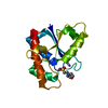
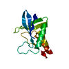
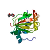
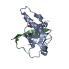
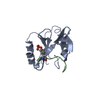
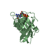
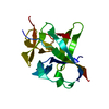
| Title | Co-crystal structure of the N-termial cGMP binding domain of Plasmodium falciparum PKG with cGMP | ||||||
 Components Components | CGMP-dependent protein kinase | ||||||
 Keywords Keywords | TRANSFERASE / Kinase / cGMP binding domain / Structural Genomics / Structural Genomics Consortium / SGC | ||||||
| Function / homology |  Function and homology information Function and homology informationcGMP-dependent protein kinase / cGMP-dependent protein kinase activity / gamete generation / extrinsic component of membrane / cAMP-dependent protein kinase activity / cAMP-dependent protein kinase complex / cGMP binding / adenylate cyclase-activating G protein-coupled receptor signaling pathway / protein phosphorylation / protein serine kinase activity ...cGMP-dependent protein kinase / cGMP-dependent protein kinase activity / gamete generation / extrinsic component of membrane / cAMP-dependent protein kinase activity / cAMP-dependent protein kinase complex / cGMP binding / adenylate cyclase-activating G protein-coupled receptor signaling pathway / protein phosphorylation / protein serine kinase activity / endoplasmic reticulum membrane / endoplasmic reticulum / ATP binding / metal ion binding / cytoplasm Similarity search - Function | ||||||
| Biological species |  | ||||||
| Method |  X-RAY DIFFRACTION / MOLECULAR REPLACEMENT / Resolution: 1.65 Å X-RAY DIFFRACTION / MOLECULAR REPLACEMENT / Resolution: 1.65 Å | ||||||
 Authors Authors | El Bakkouri, M. / Walker, J.R. / Loppnau, P. / Arrowsmith, C.H. / Edwards, A.M. / Bountra, C. / Hui, R. / Structural Genomics Consortium (SGC) | ||||||
 Citation Citation | Journal: Proc.Natl.Acad.Sci.USA / Year: 2019 Title: Structures of the cGMP-dependent protein kinase in malaria parasites reveal a unique structural relay mechanism for activation. Authors: El Bakkouri, M. / Kouidmi, I. / Wernimont, A.K. / Amani, M. / Hutchinson, A. / Loppnau, P. / Kim, J.J. / Flueck, C. / Walker, J.R. / Seitova, A. / Senisterra, G. / Kakihara, Y. / Kim, C. / ...Authors: El Bakkouri, M. / Kouidmi, I. / Wernimont, A.K. / Amani, M. / Hutchinson, A. / Loppnau, P. / Kim, J.J. / Flueck, C. / Walker, J.R. / Seitova, A. / Senisterra, G. / Kakihara, Y. / Kim, C. / Blackman, M.J. / Calmettes, C. / Baker, D.A. / Hui, R. | ||||||
| History |
|
- Structure visualization
| Structure viewer | Molecule: MolmilJmol/JSmol |
|---|
- Downloads & links
-Download
| PDBx/mmCIF format | 5e16.cif.gz | 47.4 KB | Display | PDBx/mmCIF format |
|---|---|---|---|---|
| PDB format | pdb5e16.ent.gz | 30.5 KB | Display | PDB format |
| PDBx/mmJSON format | 5e16.json.gz | Tree view | PDBx/mmJSON format | |
| Others |  Other downloads Other downloads |
-Validation report
| Arichive directory | https://data.pdbj.org/pub/pdb/validation_reports/e1/5e16ftp://data.pdbj.org/pub/pdb/validation_reports/e1/5e16 | HTTPS FTP |
|---|
-Related structure data
| Related structure data |  5dykC  5dylC  5dzcC  4myj S: Starting model for refinement C: citing same article ( |
|---|---|
| Similar structure data |


-Links
 PDBj
PDBj


- Assembly
| Deposited unit | 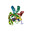
| ||||||||
|---|---|---|---|---|---|---|---|---|---|
| 1 |
| ||||||||
| Unit cell |
|
-Components
| #1: Protein | Mass: 16078.078 Da / Num. of mol.: 1 / Fragment: cGMP binding domain (UNP residues 21-162) Source method: isolated from a genetically manipulated source Source: (gene. exp.) Plasmid: pET15-MHL / Production host:  |
|---|---|
| #2: Chemical | ChemComp-PCG /   Mass: 345.205 Da / Num. of mol.: 1 / Source method: obtained synthetically / Formula: C10H12N5O7P Mass: 345.205 Da / Num. of mol.: 1 / Source method: obtained synthetically / Formula: C10H12N5O7P |
| #3: Water | ChemComp-HOH /  Mass: 18.015 Da / Num. of mol.: 104 / Source method: isolated from a natural source / Formula: H2O Mass: 18.015 Da / Num. of mol.: 104 / Source method: isolated from a natural source / Formula: H2O |
-Experimental details
-Experiment
| Experiment | Method: X-RAY DIFFRACTION / Number of used crystals: 1 |
|---|
- Sample preparation
| Crystal | Density Matthews: 1.98 Å3/Da / Density % sol: 37.94 % |
|---|---|
| Crystal grow | Temperature: 293 K / Method: vapor diffusion, sitting drop / pH: 7.5 / Details: 25 % Peg3350, 0.2 M NaCl, 0.1 M HEPES, pH 7.5 |
-Data collection
| Diffraction | Mean temperature: 100 K | |||||||||||||||||||||||||||||||||||||||||||||||||||||||||||||||||||||||||||||||||||||||||||||||||||||||||||||||||||||||||||||||||||||||||||||||||||||||||||||||||||||||||||||||||||||||||||||
|---|---|---|---|---|---|---|---|---|---|---|---|---|---|---|---|---|---|---|---|---|---|---|---|---|---|---|---|---|---|---|---|---|---|---|---|---|---|---|---|---|---|---|---|---|---|---|---|---|---|---|---|---|---|---|---|---|---|---|---|---|---|---|---|---|---|---|---|---|---|---|---|---|---|---|---|---|---|---|---|---|---|---|---|---|---|---|---|---|---|---|---|---|---|---|---|---|---|---|---|---|---|---|---|---|---|---|---|---|---|---|---|---|---|---|---|---|---|---|---|---|---|---|---|---|---|---|---|---|---|---|---|---|---|---|---|---|---|---|---|---|---|---|---|---|---|---|---|---|---|---|---|---|---|---|---|---|---|---|---|---|---|---|---|---|---|---|---|---|---|---|---|---|---|---|---|---|---|---|---|---|---|---|---|---|---|---|---|---|---|---|
| Diffraction source | Source: ROTATING ANODE / Type: RIGAKU FR-E SUPERBRIGHT / Wavelength: 1.54 Å | |||||||||||||||||||||||||||||||||||||||||||||||||||||||||||||||||||||||||||||||||||||||||||||||||||||||||||||||||||||||||||||||||||||||||||||||||||||||||||||||||||||||||||||||||||||||||||||
| Detector | Type: RIGAKU SATURN A200 / Detector: CCD / Date: Jul 2, 2015 | |||||||||||||||||||||||||||||||||||||||||||||||||||||||||||||||||||||||||||||||||||||||||||||||||||||||||||||||||||||||||||||||||||||||||||||||||||||||||||||||||||||||||||||||||||||||||||||
| Radiation | Protocol: SINGLE WAVELENGTH / Monochromatic (M) / Laue (L): M / Scattering type: x-ray | |||||||||||||||||||||||||||||||||||||||||||||||||||||||||||||||||||||||||||||||||||||||||||||||||||||||||||||||||||||||||||||||||||||||||||||||||||||||||||||||||||||||||||||||||||||||||||||
| Radiation wavelength | Wavelength: 1.54 Å / Relative weight: 1 | |||||||||||||||||||||||||||||||||||||||||||||||||||||||||||||||||||||||||||||||||||||||||||||||||||||||||||||||||||||||||||||||||||||||||||||||||||||||||||||||||||||||||||||||||||||||||||||
| Reflection | Resolution: 1.65→50 Å / Num. obs: 15745 / % possible obs: 99.7 % / Redundancy: 7.4 % / Rmerge(I) obs: 0.043 / Rpim(I) all: 0.017 / Rrim(I) all: 0.046 / Χ2: 1.587 / Net I/av σ(I): 59.607 / Net I/σ(I): 19.4 / Num. measured all: 116330 | |||||||||||||||||||||||||||||||||||||||||||||||||||||||||||||||||||||||||||||||||||||||||||||||||||||||||||||||||||||||||||||||||||||||||||||||||||||||||||||||||||||||||||||||||||||||||||||
| Reflection shell | Diffraction-ID: 1 / Rejects: _
|
- Processing
| Software |
| |||||||||||||||||||||||||||||||||||||||||||||||||||||||||||||||||||||||||||
|---|---|---|---|---|---|---|---|---|---|---|---|---|---|---|---|---|---|---|---|---|---|---|---|---|---|---|---|---|---|---|---|---|---|---|---|---|---|---|---|---|---|---|---|---|---|---|---|---|---|---|---|---|---|---|---|---|---|---|---|---|---|---|---|---|---|---|---|---|---|---|---|---|---|---|---|---|
| Refinement | Method to determine structure: MOLECULAR REPLACEMENT Starting model: 4MYJ 4myj Resolution: 1.65→46.18 Å / Cor.coef. Fo:Fc: 0.962 / Cor.coef. Fo:Fc free: 0.956 / SU B: 2.358 / SU ML: 0.081 / Cross valid method: THROUGHOUT / σ(F): 0 / ESU R: 0.117 / ESU R Free: 0.111 / Stereochemistry target values: MAXIMUM LIKELIHOOD Details: HYDROGENS HAVE BEEN ADDED IN THE RIDING POSITIONS U VALUES : REFINED INDIVIDUALLY
| |||||||||||||||||||||||||||||||||||||||||||||||||||||||||||||||||||||||||||
| Solvent computation | Ion probe radii: 0.8 Å / Shrinkage radii: 0.8 Å / VDW probe radii: 1.2 Å / Solvent model: MASK | |||||||||||||||||||||||||||||||||||||||||||||||||||||||||||||||||||||||||||
| Displacement parameters | Biso max: 88.65 Å2 / Biso mean: 27.091 Å2 / Biso min: 14.96 Å2
| |||||||||||||||||||||||||||||||||||||||||||||||||||||||||||||||||||||||||||
| Refinement step | Cycle: final / Resolution: 1.65→46.18 Å
| |||||||||||||||||||||||||||||||||||||||||||||||||||||||||||||||||||||||||||
| Refine LS restraints |
| |||||||||||||||||||||||||||||||||||||||||||||||||||||||||||||||||||||||||||
| LS refinement shell | Resolution: 1.649→1.692 Å / Total num. of bins used: 20
|
