| 登録情報 | データベース: PDB / ID: 5dqn
|
|---|
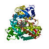
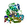
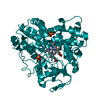
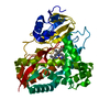
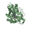
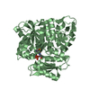
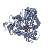
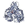
| タイトル | Polyethylene 600-bound form of P450 CYP125A3 mutant from Myobacterium Smegmatis - W83Y |
|---|
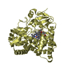
 要素 要素 | Cytochrome P450 CYP125 |
|---|
 キーワード キーワード | OXIDOREDUCTASE / CHOLESTEROL METABOLISM |
|---|
| 機能・相同性 |  機能・相同性情報 機能・相同性情報
cholest-4-en-3-one 26-monooxygenase [(25S)-3-oxocholest-4-en-26-oate forming] / cholest-4-en-3-one 26-monooxygenase activity / steroid hydroxylase activity / cholesterol catabolic process / iron ion binding / heme binding類似検索 - 分子機能 Cytochrome P450, B-class / Cytochrome p450 / Cytochrome P450 / Cytochrome P450 / Cytochrome P450 superfamily / Cytochrome P450 / Orthogonal Bundle / Mainly Alpha類似検索 - ドメイン・相同性 CITRIC ACID / PROTOPORPHYRIN IX CONTAINING FE / PHOSPHATE ION / Steroid C26-monooxygenase / Steroid C26-monooxygenase類似検索 - 構成要素 |
|---|
| 生物種 |  Mycobacterium smegmatis (バクテリア) Mycobacterium smegmatis (バクテリア) |
|---|
| 手法 |  X線回折 / シンクロトロン / 分子置換 / 解像度: 2.262 Å X線回折 / シンクロトロン / 分子置換 / 解像度: 2.262 Å |
|---|
 データ登録者 データ登録者 | Ortiz de Montellano, P.J. / Frank, D.J. / Waddling, C.A. |
|---|
| 資金援助 |  米国, 1件 米国, 1件 | 組織 | 認可番号 | 国 |
|---|
| National Institutes of Health/National Institute Of Allergy and Infectious Diseases (NIH/NIAID) | AI074824 | 米国 |
|
|---|
 引用 引用 | ジャーナル: Biochemistry / 年: 2015
タイトル: Cytochrome P450 125A4, the Third Cholesterol C-26 Hydroxylase from Mycobacterium smegmatis.
著者: Frank, D.J. / Waddling, C.A. / La, M. / Ortiz de Montellano, P.R. |
|---|
| 履歴 | | 登録 | 2015年9月15日 | 登録サイト: RCSB / 処理サイト: RCSB |
|---|
| 改定 1.0 | 2015年11月18日 | Provider: repository / タイプ: Initial release |
|---|
| 改定 1.1 | 2015年12月9日 | Group: Database references |
|---|
| 改定 1.2 | 2017年9月20日 | Group: Author supporting evidence / Database references / Derived calculations
カテゴリ: citation / pdbx_audit_support / pdbx_struct_oper_list
Item: _citation.journal_id_CSD / _pdbx_audit_support.funding_organization / _pdbx_struct_oper_list.symmetry_operation |
|---|
| 改定 1.3 | 2019年12月11日 | Group: Author supporting evidence / カテゴリ: pdbx_audit_support / Item: _pdbx_audit_support.funding_organization |
|---|
| 改定 1.4 | 2023年9月27日 | Group: Data collection / Database references / Refinement description
カテゴリ: chem_comp_atom / chem_comp_bond ...chem_comp_atom / chem_comp_bond / database_2 / pdbx_initial_refinement_model
Item: _database_2.pdbx_DOI / _database_2.pdbx_database_accession |
|---|
|
|---|
 ムービー
ムービー コントローラー


 データを開く
 基本情報
基本情報 構造の表示
構造の表示 ダウンロードとリンク
 その他のダウンロード

 PDBj
PDBj
 集合体
集合体


 分子量: 616.487 Da / 分子数: 1 / 由来タイプ: 合成 / 式: C34H32FeN4O4
 分子量: 94.971 Da / 分子数: 2 / 由来タイプ: 合成 / 式: PO4
 分子量: 192.124 Da / 分子数: 1 / 由来タイプ: 合成 / 式: C6H8O7
 分子量: 238.278 Da / 分子数: 1 / 由来タイプ: 合成 / 式: C10H22O6 / コメント: 沈殿剤*YM
 試料調製
試料調製 解析
解析