 Movie
Movie Controller


+ Open data

- Basic information
| Entry | Database: PDB / ID: 5dp1 | ||||||
|---|---|---|---|---|---|---|---|
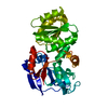
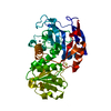
| Title | Crystal structure of CurK enoyl reductase | ||||||
 Components Components | CurK | ||||||
 Keywords Keywords | OXIDOREDUCTASE / Polyketide synthase / enoyl reductase | ||||||
| Function / homology |  Function and homology information Function and homology informationDIM/DIP cell wall layer assembly / fatty acid synthase activity / phosphopantetheine binding / fatty acid biosynthetic process / oxidoreductase activity / plasma membrane / cytoplasm Similarity search - Function | ||||||
| Biological species |  Moorea producens 3L (bacteria) Moorea producens 3L (bacteria) | ||||||
| Method |  X-RAY DIFFRACTION / SYNCHROTRON / MOLECULAR REPLACEMENT / Resolution: 1.85 Å X-RAY DIFFRACTION / SYNCHROTRON / MOLECULAR REPLACEMENT / Resolution: 1.85 Å | ||||||
 Authors Authors | Khare, D. / Smith, J.L. | ||||||
| Funding support |  United States, 1items United States, 1items
| ||||||
 Citation Citation | Journal: Structure / Year: 2015 Title: Structural Basis for Cyclopropanation by a Unique Enoyl-Acyl Carrier Protein Reductase. Authors: Khare, D. / Hale, W.A. / Tripathi, A. / Gu, L. / Sherman, D.H. / Gerwick, W.H. / Hakansson, K. / Smith, J.L. | ||||||
| History |
|
- Structure visualization
| Structure viewer | Molecule: MolmilJmol/JSmol |
|---|
- Downloads & links
-Download
| PDBx/mmCIF format | 5dp1.cif.gz | 150.2 KB | Display | PDBx/mmCIF format |
|---|---|---|---|---|
| PDB format | pdb5dp1.ent.gz | 117.2 KB | Display | PDB format |
| PDBx/mmJSON format | 5dp1.json.gz | Tree view | PDBx/mmJSON format | |
| Others |  Other downloads Other downloads |
-Validation report
| Summary document | 5dp1_validation.pdf.gz | 450.1 KB | Display | wwPDB validaton report |
|---|---|---|---|---|
| Full document | 5dp1_full_validation.pdf.gz | 451.1 KB | Display | |
| Data in XML | 5dp1_validation.xml.gz | 16.8 KB | Display | |
| Data in CIF | 5dp1_validation.cif.gz | 24.2 KB | Display | |
| Arichive directory | https://data.pdbj.org/pub/pdb/validation_reports/dp/5dp1ftp://data.pdbj.org/pub/pdb/validation_reports/dp/5dp1 | HTTPS FTP |
-Related structure data
| Related structure data | 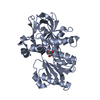 5dovC 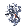 5dozC 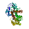 5dp2C  2vz9S C: citing same article ( S: Starting model for refinement |
|---|---|
| Similar structure data |


-Links
 PDBj
PDBj


- Assembly
| Deposited unit | 
| ||||||||||||
|---|---|---|---|---|---|---|---|---|---|---|---|---|---|
| 1 |
| ||||||||||||
| Unit cell |
| ||||||||||||
| Components on special symmetry positions |
|
-Components
| #1: Protein | Mass: 39471.758 Da / Num. of mol.: 1 / Fragment: UNP residues 1482-1815 Source method: isolated from a genetically manipulated source Source: (gene. exp.) Moorea producens 3L (bacteria) / Gene: LYNGBM3L_74450Production host: References: UniProt: F4Y425 | ||||
|---|---|---|---|---|---|
| #2: Chemical | ChemComp-GOL /   Mass: 92.094 Da / Num. of mol.: 4 / Source method: obtained synthetically / Formula: C3H8O3 Mass: 92.094 Da / Num. of mol.: 4 / Source method: obtained synthetically / Formula: C3H8O3#3: Chemical |   Mass: 94.971 Da / Num. of mol.: 2 / Source method: obtained synthetically / Formula: PO4 Mass: 94.971 Da / Num. of mol.: 2 / Source method: obtained synthetically / Formula: PO4#4: Water | ChemComp-HOH / |  Mass: 18.015 Da / Num. of mol.: 230 / Source method: isolated from a natural source / Formula: H2O Mass: 18.015 Da / Num. of mol.: 230 / Source method: isolated from a natural source / Formula: H2O |
-Experimental details
-Experiment
| Experiment | Method: X-RAY DIFFRACTION |
|---|
- Sample preparation
| Crystal | Density Matthews: 2.41 Å3/Da / Density % sol: 48.9 % |
|---|---|
| Crystal grow | Temperature: 293.15 K / Method: vapor diffusion, sitting drop Details: 1.2 M NaH2PO4, 0.8 M K2HPO4, 0.2 M Li2SO4 and 0.1 M CAPS pH 10.5 |
-Data collection
| Diffraction | Mean temperature: 100 K |
|---|---|
| Diffraction source | Source: SYNCHROTRON / Site: APS / Beamline: 23-ID-B / Wavelength: 1.0332 Å |
| Detector | Type: MARMOSAIC 300 mm CCD / Detector: CCD / Date: Aug 19, 2010 |
| Radiation | Protocol: SINGLE WAVELENGTH / Monochromatic (M) / Laue (L): M / Scattering type: x-ray |
| Radiation wavelength | Wavelength: 1.0332 Å / Relative weight: 1 |
| Reflection | Resolution: 1.85→24.3 Å / Num. obs: 32609 / % possible obs: 99 % / Redundancy: 3.8 % / Rmerge(I) obs: 0.059 / Net I/σ(I): 21 |
- Processing
| Software |
| ||||||||||||||||||||||||||||||||||||||||||||||||||||||||||||||||||||||||||||||||||||||||||||||||||||||||||||||||||||||||||||||||||||||||||||||||||||||||||||||||||||||||||||||||||||||
|---|---|---|---|---|---|---|---|---|---|---|---|---|---|---|---|---|---|---|---|---|---|---|---|---|---|---|---|---|---|---|---|---|---|---|---|---|---|---|---|---|---|---|---|---|---|---|---|---|---|---|---|---|---|---|---|---|---|---|---|---|---|---|---|---|---|---|---|---|---|---|---|---|---|---|---|---|---|---|---|---|---|---|---|---|---|---|---|---|---|---|---|---|---|---|---|---|---|---|---|---|---|---|---|---|---|---|---|---|---|---|---|---|---|---|---|---|---|---|---|---|---|---|---|---|---|---|---|---|---|---|---|---|---|---|---|---|---|---|---|---|---|---|---|---|---|---|---|---|---|---|---|---|---|---|---|---|---|---|---|---|---|---|---|---|---|---|---|---|---|---|---|---|---|---|---|---|---|---|---|---|---|---|---|
| Refinement | Method to determine structure: MOLECULAR REPLACEMENT Starting model: 2VZ9 Resolution: 1.85→24.3 Å / Cor.coef. Fo:Fc: 0.965 / Cor.coef. Fo:Fc free: 0.944 / SU B: 6.738 / SU ML: 0.099 / Cross valid method: THROUGHOUT / ESU R: 0.136 / ESU R Free: 0.128 / Stereochemistry target values: MAXIMUM LIKELIHOOD
| ||||||||||||||||||||||||||||||||||||||||||||||||||||||||||||||||||||||||||||||||||||||||||||||||||||||||||||||||||||||||||||||||||||||||||||||||||||||||||||||||||||||||||||||||||||||
| Solvent computation | Ion probe radii: 0.8 Å / Shrinkage radii: 0.8 Å / VDW probe radii: 1.2 Å / Solvent model: MASK | ||||||||||||||||||||||||||||||||||||||||||||||||||||||||||||||||||||||||||||||||||||||||||||||||||||||||||||||||||||||||||||||||||||||||||||||||||||||||||||||||||||||||||||||||||||||
| Displacement parameters | Biso mean: 36.418 Å2
| ||||||||||||||||||||||||||||||||||||||||||||||||||||||||||||||||||||||||||||||||||||||||||||||||||||||||||||||||||||||||||||||||||||||||||||||||||||||||||||||||||||||||||||||||||||||
| Refinement step | Cycle: 1 / Resolution: 1.85→24.3 Å
| ||||||||||||||||||||||||||||||||||||||||||||||||||||||||||||||||||||||||||||||||||||||||||||||||||||||||||||||||||||||||||||||||||||||||||||||||||||||||||||||||||||||||||||||||||||||
| Refine LS restraints |
|