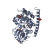
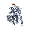
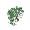
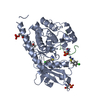
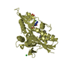
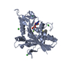
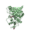
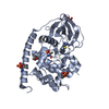
Entry Database : PDB / ID : 5do4Title Thrombin-RNA aptamer complex RNA (25-MER) Thrombin heavy chain Thrombin light chain Keywords / / / / / Function / homology Function Domain/homology Component
/ / / / / / / / / / / / / / / / / / / / / / / / / / / / / / / / / / / / / / / / / / / / / / / / / / / / / / / / / / / / / / / / / / / / / / / / / / / / / / / / / / / / / / / / / / / / / / / / / / / / / / / / / / / / / / / / / Biological species Homo sapiens (human)unidentified (others) Method / / / Resolution : 1.859 Å Authors Pallan, P.S. / Egli, M. Journal : Nucleic Acids Res. / Year : 2016Title : Evoking picomolar binding in RNA by a single phosphorodithioate linkage.Authors: Abeydeera, N.D. / Egli, M. / Cox, N. / Mercier, K. / Conde, J.N. / Pallan, P.S. / Mizurini, D.M. / Sierant, M. / Hibti, F.E. / Hassell, T. / Wang, T. / Liu, F.W. / Liu, H.M. / Martinez, C. / ... Authors : Abeydeera, N.D. / Egli, M. / Cox, N. / Mercier, K. / Conde, J.N. / Pallan, P.S. / Mizurini, D.M. / Sierant, M. / Hibti, F.E. / Hassell, T. / Wang, T. / Liu, F.W. / Liu, H.M. / Martinez, C. / Sood, A.K. / Lybrand, T.P. / Frydman, C. / Monteiro, R.Q. / Gomer, R.H. / Nawrot, B. / Yang, X. History Deposition Sep 10, 2015 Deposition site / Processing site Revision 1.0 Sep 14, 2016 Provider / Type Revision 1.1 Oct 12, 2016 Group Revision 1.2 Oct 26, 2016 Group Revision 1.3 Jul 29, 2020 Group / Derived calculations / Structure summaryCategory chem_comp / entity ... chem_comp / entity / pdbx_chem_comp_identifier / pdbx_entity_nonpoly / pdbx_struct_conn_angle / pdbx_struct_oper_list / struct_conn / struct_site / struct_site_gen Item _chem_comp.name / _chem_comp.type ... _chem_comp.name / _chem_comp.type / _entity.pdbx_description / _pdbx_entity_nonpoly.name / _pdbx_struct_conn_angle.ptnr1_auth_seq_id / _pdbx_struct_conn_angle.ptnr3_auth_seq_id / _pdbx_struct_conn_angle.value / _pdbx_struct_oper_list.symmetry_operation / _struct_conn.conn_type_id / _struct_conn.id / _struct_conn.pdbx_dist_value / _struct_conn.pdbx_leaving_atom_flag / _struct_conn.pdbx_role / _struct_conn.ptnr1_auth_asym_id / _struct_conn.ptnr1_auth_comp_id / _struct_conn.ptnr1_auth_seq_id / _struct_conn.ptnr1_label_asym_id / _struct_conn.ptnr1_label_atom_id / _struct_conn.ptnr1_label_comp_id / _struct_conn.ptnr1_label_seq_id / _struct_conn.ptnr2_auth_asym_id / _struct_conn.ptnr2_auth_comp_id / _struct_conn.ptnr2_auth_seq_id / _struct_conn.ptnr2_label_asym_id / _struct_conn.ptnr2_label_atom_id / _struct_conn.ptnr2_label_comp_id / _struct_conn.ptnr2_label_seq_id Description / Provider / Type Revision 1.4 Sep 27, 2023 Group Data collection / Database references ... Data collection / Database references / Refinement description / Structure summary Category chem_comp / chem_comp_atom ... chem_comp / chem_comp_atom / chem_comp_bond / database_2 / pdbx_initial_refinement_model Item / _database_2.pdbx_DOI / _database_2.pdbx_database_accessionRevision 1.5 Oct 30, 2024 Group / Category / pdbx_modification_feature
Show all Show less
 Movie
Movie Controller


 Open data
 Basic information
 Components
 Keywords
 Function and homology information
 Homo sapiens (human)
 X-RAY DIFFRACTION /
  Authors
 Citation
 Structure visualization
 Downloads & links
 Other downloads
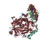


 PDBj
PDBj


 Assembly
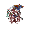

 Type: D-saccharide, beta linking / Mass: 221.208 Da / Num. of mol.: 1


 Type: peptide-like, Peptide-like / Class: Inhibitor / Mass: 453.986 Da / Num. of mol.: 1 / Source method: obtained synthetically / Formula: C21H34ClN6O3 / References: D-Phe-Pro-Arg-CH2Cl
 Mass: 62.068 Da / Num. of mol.: 4 / Source method: obtained synthetically / Formula: C2H6O2
 Mass: 92.094 Da / Num. of mol.: 2 / Source method: obtained synthetically / Formula: C3H8O3
 Mass: 40.078 Da / Num. of mol.: 1 / Source method: obtained synthetically / Formula: Ca
 Mass: 118.174 Da / Num. of mol.: 1 / Source method: obtained synthetically / Formula: C6H14O2 / Comment: precipitant*YM
 Mass: 24.305 Da / Num. of mol.: 1 / Source method: obtained synthetically / Formula: Mg
 Sample preparation
 / Beamline: 21-ID-G / Wavelength: 0.97857 Å
 Processing