[English] 日本語
Yorodumi
- PDB-5dl9: Structure of Tetragonal Lysozyme in complex with Iodine solved by... -

+
Open data


ID or keywords:

Loading...

-
Basic information

Entry
Database: PDB / ID: 5dl9
TitleStructure of Tetragonal Lysozyme in complex with Iodine solved by UWO Students
ComponentsLysozyme C
KeywordsHYDROLASE / Glycoside Hydrolase / Enzyme
Function / homology
Function and homology information


Antimicrobial peptides / Neutrophil degranulation / beta-N-acetylglucosaminidase activity / cell wall macromolecule catabolic process / lysozyme / lysozyme activity / killing of cells of another organism / defense response to Gram-negative bacterium / defense response to Gram-positive bacterium / defense response to bacterium ...Antimicrobial peptides / Neutrophil degranulation / beta-N-acetylglucosaminidase activity / cell wall macromolecule catabolic process / lysozyme / lysozyme activity / killing of cells of another organism / defense response to Gram-negative bacterium / defense response to Gram-positive bacterium / defense response to bacterium / endoplasmic reticulum / extracellular space / identical protein binding / cytoplasm
Similarity search - Function
Lysozyme - #10 / Glycoside hydrolase, family 22, lysozyme / Glycoside hydrolase family 22 domain / Glycosyl hydrolases family 22 (GH22) domain signature. / Glycoside hydrolase, family 22 / C-type lysozyme/alpha-lactalbumin family / Glycosyl hydrolases family 22 (GH22) domain profile. / Alpha-lactalbumin / lysozyme C / Lysozyme / Lysozyme-like domain superfamily ...Lysozyme - #10 / Glycoside hydrolase, family 22, lysozyme / Glycoside hydrolase family 22 domain / Glycosyl hydrolases family 22 (GH22) domain signature. / Glycoside hydrolase, family 22 / C-type lysozyme/alpha-lactalbumin family / Glycosyl hydrolases family 22 (GH22) domain profile. / Alpha-lactalbumin / lysozyme C / Lysozyme / Lysozyme-like domain superfamily / Orthogonal Bundle / Mainly Alpha
Similarity search - Domain/homology
ACETATE ION / IODIDE ION / Lysozyme C
Similarity search - Component
Biological speciesGallus gallus (chicken)
MethodX-RAY DIFFRACTION / SYNCHROTRON / SAD / Resolution: 1.38 Å
AuthorsBednarski, R. / Cirricione, N. / Greco, A. / Hodgson, R. / Kent, S. / McGowan, J. / Notherm, B. / Patt, M. / Vue, L. / Bianchetti, C.M.
CitationJournal: To Be Published
Title: Structure of Tetragonal Lysozyme in complex with Iodine solved by UWO Students
Authors: Bednarski, R. / Cirricione, N. / Greco, A. / Hodgson, R. / Kent, S. / McGowan, J. / Notherm, B. / Patt, M. / Vue, L. / Bianchetti, C.M.
History
DepositionSep 4, 2015Deposition site: RCSB / Processing site: RCSB
Revision 1.0Sep 16, 2015Provider: repository / Type: Initial release

-
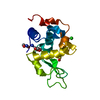
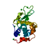
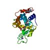
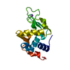
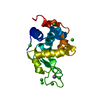
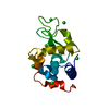
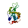
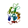
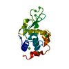
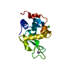
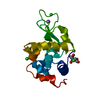
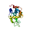
Structure visualization

Structure viewerMolecule:
MolmilJmol/JSmol

Downloads & links

-
Assembly

Deposited unit
A: Lysozyme C
hetero molecules


Theoretical massNumber of molelcules
Total (without water)15,45615
Polymers14,3311
Non-polymers1,12514
Water1,76598
1


  • Idetical with deposited unit
  • defined by author&software
TypeNameSymmetry operationNumber
identity operation1_555x,y,z1
MethodPISA
Unit cell
Length a, b, c (Å)79.459, 79.459, 37.042
Angle α, β, γ (deg.)90.000, 90.000, 90.000
Int Tables number96
Space group name H-MP43212
Components on special symmetry positions
IDModelComponents
11A-352-

HOH

21A-398-

HOH

-
Components

#1: Protein Lysozyme C / 1 / 4-beta-N-acetylmuramidase C / Allergen Gal d IV


Mass: 14331.160 Da / Num. of mol.: 1
Source method: isolated from a genetically manipulated source
Source: (gene. exp.) Gallus gallus (chicken) / Gene: LYZ / Production host: Escherichia coli (E. coli) / References: UniProt: P00698, lysozyme
#2: Chemical
ChemComp-IOD / IODIDE ION / Iodide


Mass: 126.904 Da / Num. of mol.: 4 / Source method: obtained synthetically / Formula: I
#3: Chemical
ChemComp-EDO / 1,2-ETHANEDIOL / ETHYLENE GLYCOL / Ethylene glycol


Mass: 62.068 Da / Num. of mol.: 9 / Source method: obtained synthetically / Formula: C2H6O2
#4: Chemical ChemComp-ACT / ACETATE ION / Acetate


Mass: 59.044 Da / Num. of mol.: 1 / Source method: obtained synthetically / Formula: C2H3O2
#5: Water ChemComp-HOH / water / Water


Mass: 18.015 Da / Num. of mol.: 98 / Source method: isolated from a natural source / Formula: H2O

-
Experimental details

-
Experiment

ExperimentMethod: X-RAY DIFFRACTION

-
Sample preparation

CrystalDensity Matthews: 2.04 Å3/Da / Density % sol: 39.7 %
Crystal growTemperature: 298 K / Method: vapor diffusion, hanging drop / pH: 4.5
Details: Protein Solution (60 mg/ml protein in 0.1M Na acetate pH 4.6) mixed in a 1:1 ratio with the Well Solution (10% NaCl buffered in 0.1 M Na acetate pH 4.5). Cryoprotected in 10% NaI, 0.1 M Na ...Details: Protein Solution (60 mg/ml protein in 0.1M Na acetate pH 4.6) mixed in a 1:1 ratio with the Well Solution (10% NaCl buffered in 0.1 M Na acetate pH 4.5). Cryoprotected in 10% NaI, 0.1 M Na acetate pH 4.5 25% ethylene glycol

-
Data collection

DiffractionMean temperature: 100 K
Diffraction sourceSource: SYNCHROTRON / Site: APS / Beamline: 21-ID-G / Wavelength: 0.97857 Å
DetectorType: MARMOSAIC 300 mm CCD / Detector: CCD / Date: Mar 8, 2015
RadiationProtocol: SINGLE WAVELENGTH / Monochromatic (M) / Laue (L): M / Scattering type: x-ray
Radiation wavelengthWavelength: 0.97857 Å / Relative weight: 1
ReflectionResolution: 1.23→50 Å / Num. obs: 31941 / % possible obs: 92 % / Redundancy: 12.5 % / Biso Wilson estimate: 11.79 Å2 / Rmerge(I) obs: 0.073 / Rpim(I) all: 0.02 / Rrim(I) all: 0.076 / Χ2: 2.388 / Net I/av σ(I): 51.357 / Net I/σ(I): 12.4 / Num. measured all: 400124
Reflection shell

Diffraction-ID: 1 / Rejects: 0

Resolution (Å)Redundancy (%)Rmerge(I) obsNum. unique allCC1/2Rpim(I) allRrim(I) allΧ2% possible all
1.23-1.254.20.7138650.7870.3480.7990.64551.6
1.25-1.275.70.70110120.8020.3020.7680.66758.8
1.27-1.37.20.62211530.8890.2350.6680.73568.2
1.3-1.328.40.56813100.910.20.6060.68576.2
1.32-1.359.10.50914480.9280.1720.540.72485.3
1.35-1.3910.40.41216470.9570.130.4330.78896.5
1.39-1.4211.50.34617160.9720.1050.3630.915100
1.42-1.4613.10.28517120.9790.0810.2971.027100
1.46-1.514.20.25316990.9860.0690.2631.142100
1.5-1.5514.20.21317270.990.0580.2211.305100
1.55-1.6114.30.17717250.9940.0480.1831.514100
1.61-1.6714.30.1517160.9940.0410.1561.8499.9
1.67-1.7514.30.13717430.9950.0370.1422.096100
1.75-1.8414.30.11517310.9960.0310.122.37899.9
1.84-1.9514.30.09717300.9970.0260.12.93100
1.95-2.114.30.08717470.9980.0240.0913.73100
2.1-2.3114.30.08617670.9970.0230.0894.745100
2.31-2.6514.30.08217750.9960.0220.0855.273100
2.65-3.3414.10.05917980.9990.0160.0614.353100
3.34-5013.20.04119200.9990.0120.0433.22199.6

-
Processing

Software
NameVersionClassification
PHENIX1.9_1692refinement
HKL-2000data reduction
HKL-2000data scaling
PHASERphasing
RefinementMethod to determine structure: SAD / Resolution: 1.38→33.573 Å / SU ML: 0.09 / Cross valid method: FREE R-VALUE / σ(F): 0 / Phase error: 15.23 / Stereochemistry target values: MLHL
Details: The data was collected to 1.23A, but the model was refined against data to 1.38A
RfactorNum. reflection% reflection
Rfree0.1843 1561 6.27 %
Rwork0.1545 23322 -
obs0.1564 24883 99.66 %
Solvent computationShrinkage radii: 0.9 Å / VDW probe radii: 1.11 Å / Solvent model: FLAT BULK SOLVENT MODEL
Displacement parametersBiso max: 57.76 Å2 / Biso mean: 15.2893 Å2 / Biso min: 5.92 Å2
Refinement stepCycle: final / Resolution: 1.38→33.573 Å
ProteinNucleic acidLigandSolventTotal
Num. atoms1001 0 44 98 1143
Biso mean--18.67 25.18 -
Num. residues----129
Refine LS restraints
Refine-IDTypeDev idealNumber
X-RAY DIFFRACTIONf_bond_d0.0121081
X-RAY DIFFRACTIONf_angle_d1.3221441
X-RAY DIFFRACTIONf_chiral_restr0.065148
X-RAY DIFFRACTIONf_plane_restr0.007188
X-RAY DIFFRACTIONf_dihedral_angle_d12.317391
LS refinement shell

Refine-ID: X-RAY DIFFRACTION / Total num. of bins used: 11

Resolution (Å)Rfactor RfreeNum. reflection RfreeRfactor RworkNum. reflection RworkNum. reflection all% reflection obs (%)
1.38-1.42460.16551370.12772056219398
1.4246-1.47550.17551390.12032060219999
1.4755-1.53460.15321400.1132084222499
1.5346-1.60440.18311400.113320872227100
1.6044-1.6890.16111400.118721082248100
1.689-1.79480.14181390.125220892228100
1.7948-1.93340.17341420.12421072249100
1.9334-2.12790.1421420.133421252267100
2.1279-2.43570.15731440.142521472291100
2.4357-3.06850.19881450.167521652310100
3.0685-33.5830.22921530.199922942447100

+
About Yorodumi

-
News

-
Feb 9, 2022. New format data for meta-information of EMDB entries

New format data for meta-information of EMDB entries

  • Version 3 of the EMDB header file is now the official format.
  • The previous official version 1.9 will be removed from the archive.

Related info.:EMDB header

External links:wwPDB to switch to version 3 of the EMDB data model

-
Aug 12, 2020. Covid-19 info

Covid-19 info

URL: https://pdbj.org/emnavi/covid19.php

New page: Covid-19 featured information page in EM Navigator.

Related info.:Covid-19 info / Mar 5, 2020. Novel coronavirus structure data

+
Mar 5, 2020. Novel coronavirus structure data

Novel coronavirus structure data

Related info.:Yorodumi Speices / Aug 12, 2020. Covid-19 info

External links:COVID-19 featured content - PDBj / Molecule of the Month (242):Coronavirus Proteases

+
Jan 31, 2019. EMDB accession codes are about to change! (news from PDBe EMDB page)

EMDB accession codes are about to change! (news from PDBe EMDB page)

  • The allocation of 4 digits for EMDB accession codes will soon come to an end. Whilst these codes will remain in use, new EMDB accession codes will include an additional digit and will expand incrementally as the available range of codes is exhausted. The current 4-digit format prefixed with “EMD-” (i.e. EMD-XXXX) will advance to a 5-digit format (i.e. EMD-XXXXX), and so on. It is currently estimated that the 4-digit codes will be depleted around Spring 2019, at which point the 5-digit format will come into force.
  • The EM Navigator/Yorodumi systems omit the EMD- prefix.

Related info.:Q: What is EMD? / ID/Accession-code notation in Yorodumi/EM Navigator

External links:EMDB Accession Codes are Changing Soon! / Contact to PDBj

+
Jul 12, 2017. Major update of PDB

Major update of PDB

  • wwPDB released updated PDB data conforming to the new PDBx/mmCIF dictionary.
  • This is a major update changing the version number from 4 to 5, and with Remediation, in which all the entries are updated.
  • In this update, many items about electron microscopy experimental information are reorganized (e.g. em_software).
  • Now, EM Navigator and Yorodumi are based on the updated data.

External links:wwPDB Remediation / Enriched Model Files Conforming to OneDep Data Standards Now Available in the PDB FTP Archive

-
Yorodumi

Thousand views of thousand structures

  • Yorodumi is a browser for structure data from EMDB, PDB, SASBDB, etc.
  • This page is also the successor to EM Navigator detail page, and also detail information page/front-end page for Omokage search.
  • The word "yorodu" (or yorozu) is an old Japanese word meaning "ten thousand". "mi" (miru) is to see.

Related info.:EMDB / PDB / SASBDB / Comparison of 3 databanks / Yorodumi Search / Aug 31, 2016. New EM Navigator & Yorodumi / Yorodumi Papers / Jmol/JSmol / Function and homology information / Changes in new EM Navigator and Yorodumi

Read more