 Movie
Movie Controller

[English] 日本語
 Yorodumi
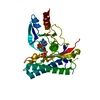
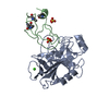
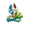
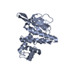
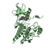
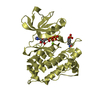
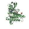
- PDB-5djg: Structure of M. tuberculosis CysQ, a PAP phosphatase with PAP, Mg... -
+ Open data

- Basic information
| Entry | Database: PDB / ID: 5djg | ||||||
|---|---|---|---|---|---|---|---|
| Title | Structure of M. tuberculosis CysQ, a PAP phosphatase with PAP, Mg, and Li bound | ||||||
 Components Components | 3'-phosphoadenosine 5'-phosphate phosphatase | ||||||
 Keywords Keywords | HYDROLASE / CysQ / PAP phosphatase / PAP / lithium | ||||||
| Function / homology |  Function and homology information Function and homology informationSulfate assimilation / inositol monophosphate phosphatase activity / 3'(2'),5'-bisphosphate nucleotidase / 3'(2'),5'-bisphosphate nucleotidase activity / inositol-phosphate phosphatase / 3'-phosphoadenosine 5'-phosphosulfate metabolic process / inositol monophosphate 1-phosphatase activity / sulfate assimilation / fructose-bisphosphatase / fructose 1,6-bisphosphate 1-phosphatase activity ...Sulfate assimilation / inositol monophosphate phosphatase activity / 3'(2'),5'-bisphosphate nucleotidase / 3'(2'),5'-bisphosphate nucleotidase activity / inositol-phosphate phosphatase / 3'-phosphoadenosine 5'-phosphosulfate metabolic process / inositol monophosphate 1-phosphatase activity / sulfate assimilation / fructose-bisphosphatase / fructose 1,6-bisphosphate 1-phosphatase activity / cobalt ion binding / manganese ion binding / magnesium ion binding / metal ion binding / plasma membrane Similarity search - Function | ||||||
| Biological species |   Mycobacterium tuberculosis (bacteria) Mycobacterium tuberculosis (bacteria) | ||||||
| Method |  X-RAY DIFFRACTION / SYNCHROTRON / MOLECULAR REPLACEMENT / Resolution: 1.951 Å X-RAY DIFFRACTION / SYNCHROTRON / MOLECULAR REPLACEMENT / Resolution: 1.951 Å | ||||||
 Authors Authors | Fisher, A.J. / Erickson, A.I. | ||||||
 Citation Citation | Journal: Biochemistry / Year: 2015 Title: Crystal Structures of Mycobacterium tuberculosis CysQ, with Substrate and Products Bound. Authors: Erickson, A.I. / Sarsam, R.D. / Fisher, A.J. | ||||||
| History |
|
- Structure visualization
| Structure viewer | Molecule: MolmilJmol/JSmol |
|---|
- Downloads & links
-Download
| PDBx/mmCIF format | 5djg.cif.gz | 73.9 KB | Display | PDBx/mmCIF format |
|---|---|---|---|---|
| PDB format | pdb5djg.ent.gz | 51.1 KB | Display | PDB format |
| PDBx/mmJSON format | 5djg.json.gz | Tree view | PDBx/mmJSON format | |
| Others |  Other downloads Other downloads |
-Validation report
| Arichive directory | https://data.pdbj.org/pub/pdb/validation_reports/dj/5djgftp://data.pdbj.org/pub/pdb/validation_reports/dj/5djg | HTTPS FTP |
|---|
-Related structure data
| Related structure data |  5djfSC  5djhC  5djiC  5djjC  5djkC S: Starting model for refinement C: citing same article ( |
|---|---|
| Similar structure data |


-Links
 PDBj
PDBj

- Assembly
| Deposited unit | 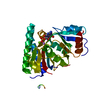
| ||||||||
|---|---|---|---|---|---|---|---|---|---|
| 1 |
| ||||||||
| Unit cell |
|
-Components
| #1: Protein | Mass: 30746.596 Da / Num. of mol.: 1 Source method: isolated from a genetically manipulated source Source: (gene. exp.) Mycobacterium tuberculosis (bacteria) / Gene: cysQ, MT2189 / Plasmid: pET28b / Production host: References: UniProt: P9WKJ0, UniProt: P9WKJ1*PLUS, 3'(2'),5'-bisphosphate nucleotidase, fructose-bisphosphatase, inositol-phosphate phosphatase |
|---|---|
| #2: Chemical | ChemComp-MG /   Mass: 24.305 Da / Num. of mol.: 1 / Source method: obtained synthetically / Formula: Mg Mass: 24.305 Da / Num. of mol.: 1 / Source method: obtained synthetically / Formula: Mg |
| #3: Chemical | ChemComp-LI /   Mass: 6.941 Da / Num. of mol.: 1 / Source method: obtained synthetically / Formula: Li Mass: 6.941 Da / Num. of mol.: 1 / Source method: obtained synthetically / Formula: Li |
| #4: Chemical | ChemComp-A3P / 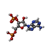  Type: RNA linking / Mass: 427.201 Da / Num. of mol.: 1 / Source method: obtained synthetically / Formula: C10H15N5O10P2 Type: RNA linking / Mass: 427.201 Da / Num. of mol.: 1 / Source method: obtained synthetically / Formula: C10H15N5O10P2 |
| #5: Water | ChemComp-HOH /  Mass: 18.015 Da / Num. of mol.: 273 / Source method: isolated from a natural source / Formula: H2O Mass: 18.015 Da / Num. of mol.: 273 / Source method: isolated from a natural source / Formula: H2O |
-Experimental details
-Experiment
| Experiment | Method: X-RAY DIFFRACTION |
|---|
- Sample preparation
| Crystal | Density Matthews: 1.94 Å3/Da / Density % sol: 36.7 % |
|---|---|
| Crystal grow | Temperature: 298.15 K / Method: vapor diffusion, sitting drop / Details: 0.2 M lithium chloride, 25% PEG8000, 1 mM PAP |
-Data collection
| Diffraction | Mean temperature: 100 K |
|---|---|
| Diffraction source | Source: SYNCHROTRON / Site: SSRL  / Beamline: BL7-1 / Wavelength: 1.12709 Å / Beamline: BL7-1 / Wavelength: 1.12709 Å |
| Detector | Type: ADSC QUANTUM 315r / Detector: CCD / Date: May 28, 2014 |
| Radiation | Monochromator: Side scattering I-beam bent single crystal, asymmetric cut 4.9650 degrees Protocol: SINGLE WAVELENGTH / Monochromatic (M) / Laue (L): M / Scattering type: x-ray |
| Radiation wavelength | Wavelength: 1.12709 Å / Relative weight: 1 |
| Reflection | Resolution: 1.951→38.151 Å / Num. all: 17281 / Num. obs: 17281 / % possible obs: 95.5 % / Observed criterion σ(F): 0 / Observed criterion σ(I): 0 / Redundancy: 3.39 % / Rmerge(I) obs: 0.071 / Net I/σ(I): 14.44 |
| Reflection shell | Resolution: 1.951→2 Å / Redundancy: 2.44 % / Rmerge(I) obs: 0.265 / Mean I/σ(I) obs: 4.43 / % possible all: 84.1 |
- Processing
| Software |
| |||||||||||||||||||||||||||||||||||||||||||||||||||||||||||||||||||||||||||||||||||||||||||
|---|---|---|---|---|---|---|---|---|---|---|---|---|---|---|---|---|---|---|---|---|---|---|---|---|---|---|---|---|---|---|---|---|---|---|---|---|---|---|---|---|---|---|---|---|---|---|---|---|---|---|---|---|---|---|---|---|---|---|---|---|---|---|---|---|---|---|---|---|---|---|---|---|---|---|---|---|---|---|---|---|---|---|---|---|---|---|---|---|---|---|---|---|
| Refinement | Method to determine structure: MOLECULAR REPLACEMENT Starting model: PDB entry 5DJF Resolution: 1.951→38.151 Å / SU ML: 0.16 / Cross valid method: FREE R-VALUE / σ(F): 0.9 / Phase error: 16.74 / Stereochemistry target values: ML
| |||||||||||||||||||||||||||||||||||||||||||||||||||||||||||||||||||||||||||||||||||||||||||
| Solvent computation | Shrinkage radii: 0.9 Å / VDW probe radii: 1.11 Å / Solvent model: FLAT BULK SOLVENT MODEL | |||||||||||||||||||||||||||||||||||||||||||||||||||||||||||||||||||||||||||||||||||||||||||
| Refinement step | Cycle: LAST / Resolution: 1.951→38.151 Å
| |||||||||||||||||||||||||||||||||||||||||||||||||||||||||||||||||||||||||||||||||||||||||||
| Refine LS restraints |
| |||||||||||||||||||||||||||||||||||||||||||||||||||||||||||||||||||||||||||||||||||||||||||
| LS refinement shell |
|
