 Movie
Movie Controller

[English] 日本語
 Yorodumi
- PDB-5di3: Crystal structure of Arl13B in complex with Arl3 of Chlamydomonas... -
+ Open data

- Basic information
| Entry | Database: PDB / ID: 5di3 | ||||||
|---|---|---|---|---|---|---|---|
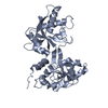
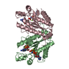
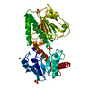
| Title | Crystal structure of Arl13B in complex with Arl3 of Chlamydomonas reinhardtii | ||||||
 Components Components |
| ||||||
 Keywords Keywords | HYDROLASE / G-protein / ADP ribosylation like protein / Complex / Guanine nucleotide exchange factor | ||||||
| Function / homology |  Function and homology information Function and homology informationciliary membrane / microtubule organizing center / spindle / protein transport / Golgi membrane / cell division / GTPase activity / GTP binding / nucleus Similarity search - Function | ||||||
| Biological species |   Chlamydomonas reinhardtii (plant) Chlamydomonas reinhardtii (plant) | ||||||
| Method |  X-RAY DIFFRACTION / SYNCHROTRON / MOLECULAR REPLACEMENT / molecular replacement / Resolution: 2.5 Å X-RAY DIFFRACTION / SYNCHROTRON / MOLECULAR REPLACEMENT / molecular replacement / Resolution: 2.5 Å | ||||||
 Authors Authors | Gotthardt, K. / Lokaj, M. / Falk, N. / Koerner, C. / Giessl, A. / Wittinghofer, A. | ||||||
 Citation Citation | Journal: Elife / Year: 2015 Title: A G-protein activation cascade from Arl13B to Arl3 and implications for ciliary targeting of lipidated proteins. Authors: Gotthardt, K. / Lokaj, M. / Koerner, C. / Falk, N. / Giel, A. / Wittinghofer, A. | ||||||
| History |
|
- Structure visualization
| Structure viewer | Molecule: MolmilJmol/JSmol |
|---|
- Downloads & links
-Download
| PDBx/mmCIF format | 5di3.cif.gz | 91.9 KB | Display | PDBx/mmCIF format |
|---|---|---|---|---|
| PDB format | pdb5di3.ent.gz | 66.6 KB | Display | PDB format |
| PDBx/mmJSON format | 5di3.json.gz | Tree view | PDBx/mmJSON format | |
| Others |  Other downloads Other downloads |
-Validation report
| Summary document | 5di3_validation.pdf.gz | 1 MB | Display | wwPDB validaton report |
|---|---|---|---|---|
| Full document | 5di3_full_validation.pdf.gz | 1 MB | Display | |
| Data in XML | 5di3_validation.xml.gz | 16.6 KB | Display | |
| Data in CIF | 5di3_validation.cif.gz | 21.3 KB | Display | |
| Arichive directory | https://data.pdbj.org/pub/pdb/validation_reports/di/5di3ftp://data.pdbj.org/pub/pdb/validation_reports/di/5di3 | HTTPS FTP |
-Related structure data
| Related structure data |  3bh6S S: Starting model for refinement |
|---|---|
| Similar structure data |


-Links
 PDBj
PDBj


- Assembly
| Deposited unit | 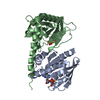
| ||||||||
|---|---|---|---|---|---|---|---|---|---|
| 1 |
| ||||||||
| Unit cell |
|
-Components
| #1: Protein | Mass: 29268.100 Da / Num. of mol.: 1 / Fragment: UNP residues 18-278 Source method: isolated from a genetically manipulated source Source: (gene. exp.) Chlamydomonas reinhardtii (plant) / Gene: ARL13, CHLREDRAFT_195529 / Production host:  | ||||
|---|---|---|---|---|---|
| #2: Protein | Mass: 20876.908 Da / Num. of mol.: 1 Source method: isolated from a genetically manipulated source Source: (gene. exp.) Chlamydomonas reinhardtii (plant) / Gene: ARL3, CHLREDRAFT_128761 / Production host: | ||||
| #3: Chemical |   Mass: 522.196 Da / Num. of mol.: 2 / Source method: obtained synthetically / Formula: C10H17N6O13P3 Mass: 522.196 Da / Num. of mol.: 2 / Source method: obtained synthetically / Formula: C10H17N6O13P3Comment: GppNHp, GMPPNP, energy-carrying molecule analogue*YM #4: Chemical |   Mass: 24.305 Da / Num. of mol.: 2 / Source method: obtained synthetically / Formula: Mg Mass: 24.305 Da / Num. of mol.: 2 / Source method: obtained synthetically / Formula: Mg#5: Water | ChemComp-HOH / |  Mass: 18.015 Da / Num. of mol.: 29 / Source method: isolated from a natural source / Formula: H2O Mass: 18.015 Da / Num. of mol.: 29 / Source method: isolated from a natural source / Formula: H2O |
-Experimental details
-Experiment
| Experiment | Method: X-RAY DIFFRACTION / Number of used crystals: 1 |
|---|
- Sample preparation
| Crystal | Density Matthews: 2.83 Å3/Da / Density % sol: 56.51 % |
|---|---|
| Crystal grow | Temperature: 293 K / Method: vapor diffusion, sitting drop / Details: Tris pH 8.5, 25 % PEG 6000 / PH range: 8.5 |
-Data collection
| Diffraction | Mean temperature: 100 K | ||||||||||||||||||||||||||||||||||||||||||||||||||||||||||||||||||||||||||||||||||||||||||
|---|---|---|---|---|---|---|---|---|---|---|---|---|---|---|---|---|---|---|---|---|---|---|---|---|---|---|---|---|---|---|---|---|---|---|---|---|---|---|---|---|---|---|---|---|---|---|---|---|---|---|---|---|---|---|---|---|---|---|---|---|---|---|---|---|---|---|---|---|---|---|---|---|---|---|---|---|---|---|---|---|---|---|---|---|---|---|---|---|---|---|---|
| Diffraction source | Source: SYNCHROTRON / Site: SLS  / Beamline: X10SA / Wavelength: 0.916 Å / Beamline: X10SA / Wavelength: 0.916 Å | ||||||||||||||||||||||||||||||||||||||||||||||||||||||||||||||||||||||||||||||||||||||||||
| Detector | Type: DECTRIS PILATUS 6M / Detector: PIXEL / Date: Feb 16, 2015 | ||||||||||||||||||||||||||||||||||||||||||||||||||||||||||||||||||||||||||||||||||||||||||
| Radiation | Monochromator: Si(111) / Protocol: SINGLE WAVELENGTH / Monochromatic (M) / Laue (L): M / Scattering type: x-ray | ||||||||||||||||||||||||||||||||||||||||||||||||||||||||||||||||||||||||||||||||||||||||||
| Radiation wavelength | Wavelength: 0.916 Å / Relative weight: 1 | ||||||||||||||||||||||||||||||||||||||||||||||||||||||||||||||||||||||||||||||||||||||||||
| Reflection | Resolution: 2.5→50 Å / Num. obs: 16944 / % possible obs: 99.9 % / Observed criterion σ(I): -3 / Redundancy: 6.4 % / Biso Wilson estimate: 60.077 Å2 / Rmerge F obs: 0.999 / Rmerge(I) obs: 0.065 / Rrim(I) all: 0.071 / Χ2: 0.98 / Net I/σ(I): 17.56 / Num. measured all: 109004 | ||||||||||||||||||||||||||||||||||||||||||||||||||||||||||||||||||||||||||||||||||||||||||
| Reflection shell | Diffraction-ID: 1 / Rejects: _
|
-Phasing
| Phasing | Method: molecular replacement |
|---|
- Processing
| Software |
| ||||||||||||||||||||||||
|---|---|---|---|---|---|---|---|---|---|---|---|---|---|---|---|---|---|---|---|---|---|---|---|---|---|
| Refinement | Method to determine structure: MOLECULAR REPLACEMENT Starting model: 3BH6 Resolution: 2.5→29.843 Å / SU ML: 0.31 / Cross valid method: FREE R-VALUE / σ(F): 1.36 / Phase error: 28.08 / Stereochemistry target values: ML
| ||||||||||||||||||||||||
| Solvent computation | Shrinkage radii: 0.9 Å / VDW probe radii: 1.11 Å / Solvent model: FLAT BULK SOLVENT MODEL | ||||||||||||||||||||||||
| Displacement parameters | Biso max: 161.06 Å2 / Biso mean: 65.9867 Å2 / Biso min: 33.73 Å2 | ||||||||||||||||||||||||
| Refinement step | Cycle: final / Resolution: 2.5→29.843 Å
|
