 Movie
Movie Controller

[English] 日本語
 Yorodumi
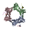
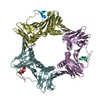
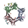
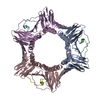
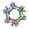
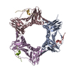
- PDB-5dai: Proliferating cell nuclear antigen homolog 1 bound to FEN-1 peptide -
+ Open data

- Basic information
| Entry | Database: PDB / ID: 5dai | ||||||
|---|---|---|---|---|---|---|---|
| Title | Proliferating cell nuclear antigen homolog 1 bound to FEN-1 peptide | ||||||
 Components Components |
| ||||||
 Keywords Keywords | TRANSFERASE / complex / PIP box binder | ||||||
| Function / homology |  Function and homology information Function and homology information5'-flap endonuclease activity / DNA replication, removal of RNA primer / 5'-3' exonuclease activity / leading strand elongation / DNA polymerase processivity factor activity / regulation of DNA replication / Hydrolases; Acting on ester bonds / DNA repair / magnesium ion binding / DNA binding / identical protein binding Similarity search - Function | ||||||
| Biological species |   Thermococcus kodakarensis (archaea)Thermococcus kodakarensis KOD1 (archaea) Thermococcus kodakarensis (archaea)Thermococcus kodakarensis KOD1 (archaea) | ||||||
| Method |  X-RAY DIFFRACTION / MOLECULAR REPLACEMENT / Resolution: 2 Å X-RAY DIFFRACTION / MOLECULAR REPLACEMENT / Resolution: 2 Å | ||||||
 Authors Authors | Ladner, J.E. / Altieri, A.S. / Kelman, Z. | ||||||
 Citation Citation | Journal: Nucleic Acids Res. / Year: 2016 Title: A small protein inhibits proliferating cell nuclear antigen by breaking the DNA clamp. Authors: Altieri, A.S. / Ladner, J.E. / Li, Z. / Robinson, H. / Sallman, Z.F. / Marino, J.P. / Kelman, Z. | ||||||
| History |
|
- Structure visualization
| Structure viewer | Molecule: MolmilJmol/JSmol |
|---|
- Downloads & links
-Download
| PDBx/mmCIF format | 5dai.cif.gz | 70.1 KB | Display | PDBx/mmCIF format |
|---|---|---|---|---|
| PDB format | pdb5dai.ent.gz | 51.4 KB | Display | PDB format |
| PDBx/mmJSON format | 5dai.json.gz | Tree view | PDBx/mmJSON format | |
| Others |  Other downloads Other downloads |
-Validation report
| Arichive directory | https://data.pdbj.org/pub/pdb/validation_reports/da/5daiftp://data.pdbj.org/pub/pdb/validation_reports/da/5dai | HTTPS FTP |
|---|
-Related structure data
| Related structure data |  5da7C  3lx1S S: Starting model for refinement C: citing same article ( |
|---|---|
| Similar structure data |


-Links
 PDBj
PDBj


- Assembly
| Deposited unit | 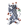
| ||||||||
|---|---|---|---|---|---|---|---|---|---|
| 1 | 
| ||||||||
| Unit cell |
|
-Components
| #1: Protein | Mass: 29099.311 Da / Num. of mol.: 1 Source method: isolated from a genetically manipulated source Source: (gene. exp.) Thermococcus kodakarensis (strain ATCC BAA-918 / JCM 12380 / KOD1) (archaea)Strain: ATCC BAA-918 / JCM 12380 / KOD1 / Gene: pcn1, TK0535 / Production host:  | ||
|---|---|---|---|
| #2: Protein/peptide | Mass: 1410.600 Da / Num. of mol.: 1 / Source method: obtained synthetically / Source: (synth.) Thermococcus kodakarensis KOD1 (archaea) / References: UniProt: Q5JGN0*PLUS | ||
| #3: Chemical | ChemComp-SO4 /   Mass: 96.063 Da / Num. of mol.: 11 / Source method: obtained synthetically / Formula: SO4 Mass: 96.063 Da / Num. of mol.: 11 / Source method: obtained synthetically / Formula: SO4#4: Water | ChemComp-HOH / |  Mass: 18.015 Da / Num. of mol.: 98 / Source method: isolated from a natural source / Formula: H2O Mass: 18.015 Da / Num. of mol.: 98 / Source method: isolated from a natural source / Formula: H2O |
-Experimental details
-Experiment
| Experiment | Method: X-RAY DIFFRACTION |
|---|
- Sample preparation
| Crystal | Density Matthews: 2.68 Å3/Da / Density % sol: 54.19 % |
|---|---|
| Crystal grow | Temperature: 298 K / Method: vapor diffusion, sitting drop / pH: 5.3 Details: Well solution: 2.8 M ammonium sulfate, 100 mM citric acid buffer pH 5.3, and 10% PEG 4000. Temp details: room temperature |
-Data collection
| Diffraction | Mean temperature: 100 K |
|---|---|
| Diffraction source | Source: ROTATING ANODE / Type: RIGAKU MICROMAX-007 / Wavelength: 1.54 Å |
| Detector | Type: RIGAKU RAXIS IV++ / Detector: IMAGE PLATE / Date: Sep 11, 2014 |
| Radiation | Protocol: SINGLE WAVELENGTH / Monochromatic (M) / Laue (L): M / Scattering type: x-ray |
| Radiation wavelength | Wavelength: 1.54 Å / Relative weight: 1 |
| Reflection | Resolution: 2→28.8 Å / Num. obs: 20143 / % possible obs: 98.2 % / Redundancy: 3.31 % / Rmerge(I) obs: 0.043 / Net I/σ(I): 11.9 |
- Processing
| Software |
| ||||||||||||||||||||||||||||||||||||||||||||||||||||||||
|---|---|---|---|---|---|---|---|---|---|---|---|---|---|---|---|---|---|---|---|---|---|---|---|---|---|---|---|---|---|---|---|---|---|---|---|---|---|---|---|---|---|---|---|---|---|---|---|---|---|---|---|---|---|---|---|---|---|
| Refinement | Method to determine structure: MOLECULAR REPLACEMENT Starting model: 3LX1 Resolution: 2→28.762 Å / SU ML: 0.3 / Cross valid method: FREE R-VALUE / σ(F): 1.98 / Phase error: 28.6 / Stereochemistry target values: ML
| ||||||||||||||||||||||||||||||||||||||||||||||||||||||||
| Solvent computation | Shrinkage radii: 0.9 Å / VDW probe radii: 1.11 Å / Solvent model: FLAT BULK SOLVENT MODEL | ||||||||||||||||||||||||||||||||||||||||||||||||||||||||
| Refinement step | Cycle: LAST / Resolution: 2→28.762 Å
| ||||||||||||||||||||||||||||||||||||||||||||||||||||||||
| Refine LS restraints |
| ||||||||||||||||||||||||||||||||||||||||||||||||||||||||
| LS refinement shell |
|
