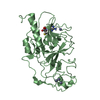
RNA polymerase II intronic transcription regulatory region sequence-specific DNA binding / RNA polymerase II transcription regulatory region sequence-specific DNA binding / sequence-specific double-stranded DNA binding / DNA-binding transcription activator activity, RNA polymerase II-specific / spermatogenesis / DNA-binding transcription factor activity, RNA polymerase II-specific / RNA polymerase II cis-regulatory region sequence-specific DNA binding / chromatin / positive regulation of transcription by RNA polymerase II / nucleoplasm ...RNA polymerase II intronic transcription regulatory region sequence-specific DNA binding / RNA polymerase II transcription regulatory region sequence-specific DNA binding / sequence-specific double-stranded DNA binding / DNA-binding transcription activator activity, RNA polymerase II-specific / spermatogenesis / DNA-binding transcription factor activity, RNA polymerase II-specific / RNA polymerase II cis-regulatory region sequence-specific DNA binding / chromatin / positive regulation of transcription by RNA polymerase II / nucleoplasm / identical protein binding / cytoplasm Similarity search - Function
Mass: 18.015 Da / Num. of mol.: 106 / Source method: isolated from a natural source / Formula: H2O
-
Experimental details
-
Experiment
Experiment
Method: X-RAY DIFFRACTION
-
Sample preparation
Crystal
Density Matthews: 1.98 Å3/Da / Density % sol: 37.93 %
Crystal grow
Temperature: 295 K / Method: vapor diffusion, sitting drop / pH: 9 Details: 7 mg/ml protein:DNA complexes were mixed at a ratio of 1:1.2 protein:DNA in 25 mM HEPES pH 7.5 and 150 mM NaCl and crystallized against 100 mM NaCl, 100 mM BICINE pH 9, and 30% PEG 550 MME. ...Details: 7 mg/ml protein:DNA complexes were mixed at a ratio of 1:1.2 protein:DNA in 25 mM HEPES pH 7.5 and 150 mM NaCl and crystallized against 100 mM NaCl, 100 mM BICINE pH 9, and 30% PEG 550 MME. Crystals grew overnight at room temperature
-
Data collection
Diffraction
Mean temperature: 93 K
Diffraction source
Source: ROTATING ANODE / Type: RIGAKU / Wavelength: 1.54 Å
In the structure databanks used in Yorodumi, some data are registered as the other names, "COVID-19 virus" and "2019-nCoV". Here are the details of the virus and the list of structure data.
Jan 31, 2019. EMDB accession codes are about to change! (news from PDBe EMDB page)
EMDB accession codes are about to change! (news from PDBe EMDB page)
The allocation of 4 digits for EMDB accession codes will soon come to an end. Whilst these codes will remain in use, new EMDB accession codes will include an additional digit and will expand incrementally as the available range of codes is exhausted. The current 4-digit format prefixed with “EMD-” (i.e. EMD-XXXX) will advance to a 5-digit format (i.e. EMD-XXXXX), and so on. It is currently estimated that the 4-digit codes will be depleted around Spring 2019, at which point the 5-digit format will come into force.
The EM Navigator/Yorodumi systems omit the EMD- prefix.
Related info.:Q: What is EMD? / ID/Accession-code notation in Yorodumi/EM Navigator
Yorodumi is a browser for structure data from EMDB, PDB, SASBDB, etc.
This page is also the successor to EM Navigator detail page, and also detail information page/front-end page for Omokage search.
The word "yorodu" (or yorozu) is an old Japanese word meaning "ten thousand". "mi" (miru) is to see.
Related info.:EMDB / PDB / SASBDB / Comparison of 3 databanks / Yorodumi Search / Aug 31, 2016. New EM Navigator & Yorodumi / Yorodumi Papers / Jmol/JSmol / Function and homology information / Changes in new EM Navigator and Yorodumi
 Movie
Movie Controller

 Yorodumi
 Open data
 Basic information
 Components
 Keywords
 Function and homology information
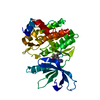
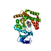
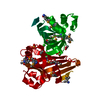
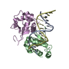
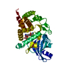
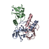
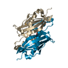
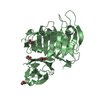
 Homo sapiens (human)
 X-RAY DIFFRACTION /
  Authors
 Citation
 Structure visualization
 Downloads & links
 Other downloads

 PDBj
PDBj


 Assembly
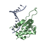


 Mass: 18.015 Da / Num. of mol.: 106 / Source method: isolated from a natural source / Formula: H2O
 Sample preparation
 Processing