 Movie
Movie Controller

[English] 日本語
 Yorodumi
- PDB-5d8h: CRYSTAL STRUCTURE OF THE BASE OF THE RIBOSOMAL P STALK FROM METHA... -
+ Open data

- Basic information
| Entry | Database: PDB / ID: 5d8h | |||||||||
|---|---|---|---|---|---|---|---|---|---|---|
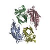
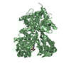
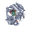
| Title | CRYSTAL STRUCTURE OF THE BASE OF THE RIBOSOMAL P STALK FROM METHANOCOCCUS JANNASCHII WITH ANTIBIOTIC THIOSTREPTON | |||||||||
 Components Components |
| |||||||||
 Keywords Keywords | RIBOSOMAL PROTEIN / ribosome / P-stalk / archaea / ANTIBIOTIC / THIOSTREPTON | |||||||||
| Function / homology |  Function and homology information Function and homology informationlarge ribosomal subunit / large ribosomal subunit rRNA binding / cytosolic large ribosomal subunit / cytoplasmic translation / defense response to bacterium / structural constituent of ribosome / translation / extracellular region Similarity search - Function | |||||||||
| Biological species |   Methanocaldococcus jannaschii (archaea) Methanocaldococcus jannaschii (archaea) Streptomyces azureus (bacteria) Streptomyces azureus (bacteria) | |||||||||
| Method |  X-RAY DIFFRACTION / SYNCHROTRON / SAD / Resolution: 2.8 Å X-RAY DIFFRACTION / SYNCHROTRON / SAD / Resolution: 2.8 Å | |||||||||
 Authors Authors | Gabdulkhakov, A.G. / Mitroshin, I.V. / Garber, M.B. | |||||||||
 Citation Citation | Journal: To Be Published Title: CRYSTAL STRUCTURE OF THE BASE OF THE RIBOSOMAL P STALK FROM METHANOCOCCUS JANNASCHII WITH ANTIBIOTIC THIOSTREPTON Authors: Gabdulkhakov, A.G. / Mitroshin, I.V. / Garber, M.B. | |||||||||
| History |
|
- Structure visualization
| Structure viewer | Molecule: MolmilJmol/JSmol |
|---|
- Downloads & links
-Download
| PDBx/mmCIF format | 5d8h.cif.gz | 144 KB | Display | PDBx/mmCIF format |
|---|---|---|---|---|
| PDB format | pdb5d8h.ent.gz | 104 KB | Display | PDB format |
| PDBx/mmJSON format | 5d8h.json.gz | Tree view | PDBx/mmJSON format | |
| Others |  Other downloads Other downloads |
-Validation report
| Summary document | 5d8h_validation.pdf.gz | 499.3 KB | Display | wwPDB validaton report |
|---|---|---|---|---|
| Full document | 5d8h_full_validation.pdf.gz | 520.6 KB | Display | |
| Data in XML | 5d8h_validation.xml.gz | 25.1 KB | Display | |
| Data in CIF | 5d8h_validation.cif.gz | 33 KB | Display | |
| Arichive directory | https://data.pdbj.org/pub/pdb/validation_reports/d8/5d8hftp://data.pdbj.org/pub/pdb/validation_reports/d8/5d8h | HTTPS FTP |
-Related structure data
| Related structure data | |
|---|---|
| Similar structure data |


-Links
 PDBj
PDBj


- Assembly
| Deposited unit | 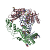
| ||||||||
|---|---|---|---|---|---|---|---|---|---|
| 1 |
| ||||||||
| Unit cell |
|
-Components
-50S ribosomal protein ... , 2 types, 2 molecules BC
| #2: Protein | Mass: 23635.162 Da / Num. of mol.: 1 / Fragment: UNP residues 9-221 / Mutation: Met changed to SeMet Source method: isolated from a genetically manipulated source Source: (gene. exp.) Methanocaldococcus jannaschii (archaea)Gene: rpl10, rplP0, MJ0509 / Plasmid: pET-11c/MjaP0NTF / Production host: |
|---|---|
| #3: Protein | Mass: 17794.695 Da / Num. of mol.: 1 Source method: isolated from a genetically manipulated source Source: (gene. exp.) Methanocaldococcus jannaschii (archaea)Gene: rpl11, MJ0373 / Plasmid: pET-11c/MjaL11 / Production host: |
-RNA chain / Protein/peptide , 2 types, 2 molecules AD
| #1: RNA chain | Mass: 23928.295 Da / Num. of mol.: 1 Source method: isolated from a genetically manipulated source Source: (gene. exp.) Methanocaldococcus jannaschii (archaea)Plasmid: pMja23S-74.UC18 / Production host: |
|---|---|
| #4: Protein/peptide |   Type: Thiopeptide / Class: Antibiotic / Mass: 1805.985 Da / Num. of mol.: 1 / Source method: isolated from a natural source Type: Thiopeptide / Class: Antibiotic / Mass: 1805.985 Da / Num. of mol.: 1 / Source method: isolated from a natural sourceDetails: Thiostrepton is a hetrocyclic thiopeptide belonging to the thiocillin family, consisting of four thiazole, one thiozoline and one piperideine rings. A modified quinoline linked to main-chain ...Details: Thiostrepton is a hetrocyclic thiopeptide belonging to the thiocillin family, consisting of four thiazole, one thiozoline and one piperideine rings. A modified quinoline linked to main-chain residue 1 and side-chain of residue 12. Post translational maturation of thiazole and oxazole containing antibiotics involves the enzymic condensation of a Cys or Ser with the alpha-carbonyl of the preceding amino acid to form a thioether or ether bond, then dehydration to form a double bond with the alpha-amino nitrogen. Thiazoline or oxazoline ring are dehydrogenated to form thiazole or oxazole rings. the pyridinyl involves the cross-linking of a Ser and a Cys-Ser pair usually separated by 7 or 8 residues along the peptide chain. The Ser residues are dehydrated to didehydroalanines, then bonded between their beta carbons. The alpha carbonyl of the Cys condenses with alpha carbon of the first Ser to form a pyridinyl ring. The ring may be mutiply dehydrogenated to form a pyridine ring with loss of the amino nitrogen of the first Ser. The amidation of Ser-17 probably does not occur by the same mechanism, oxidative cleavage of glycine, as in eukaryotes. Source: (natural) Streptomyces azureus (bacteria) / References: UniProt: P0C8P8, THIOSTREPTON |
-Non-polymers , 4 types, 83 molecules 


| #5: Chemical | ChemComp-MG /  Mass: 24.305 Da / Num. of mol.: 11 / Source method: obtained synthetically / Formula: Mg Mass: 24.305 Da / Num. of mol.: 11 / Source method: obtained synthetically / Formula: Mg#6: Chemical | ChemComp-NA /  Mass: 22.990 Da / Num. of mol.: 4 / Source method: obtained synthetically / Formula: Na Mass: 22.990 Da / Num. of mol.: 4 / Source method: obtained synthetically / Formula: Na#7: Chemical |  Mass: 122.143 Da / Num. of mol.: 3 / Source method: obtained synthetically / Formula: C4H12NO3 / Comment: pH buffer*YM Mass: 122.143 Da / Num. of mol.: 3 / Source method: obtained synthetically / Formula: C4H12NO3 / Comment: pH buffer*YM#8: Water | ChemComp-HOH / | Mass: 18.015 Da / Num. of mol.: 65 / Source method: isolated from a natural source / Formula: H2O |
|---|
-Details
| Compound details | THIOSTREPTON IS A MEMBER OF A SULPHUR-RICH HETEROCYCLIC PEPTIDES CLASS. ALL SHARE A MACROCYLIC ...THIOSTREPT |
|---|---|
| Has protein modification | Y |
-Experimental details
-Experiment
| Experiment | Method: X-RAY DIFFRACTION |
|---|
- Sample preparation
| Crystal | Density Matthews: 3.97 Å3/Da / Density % sol: 69 % |
|---|---|
| Crystal grow | Temperature: 295 K / Method: vapor diffusion, sitting drop / pH: 7.5 Details: 0.1 mM Tris-HCl, pH 7.5, 10% PEG 8000, 20% glycerol, 1 mM TCEP (tris(2-carboxyethyl)phosphine), 0.125 mM CTAB |
-Data collection
| Diffraction | Mean temperature: 100 K |
|---|---|
| Diffraction source | Source: SYNCHROTRON / Site: ESRF  / Beamline: ID23-1 / Wavelength: 0.97953 Å / Beamline: ID23-1 / Wavelength: 0.97953 Å |
| Detector | Type: DECTRIS PILATUS 6M / Detector: PIXEL / Date: Feb 13, 2015 |
| Radiation | Protocol: SINGLE WAVELENGTH / Monochromatic (M) / Laue (L): M / Scattering type: x-ray |
| Radiation wavelength | Wavelength: 0.97953 Å / Relative weight: 1 |
| Reflection | Resolution: 2.8→50 Å / Num. all: 50260 / Num. obs: 50260 / % possible obs: 99.3 % / Redundancy: 3.47 % / Rsym value: 0.061 / Net I/σ(I): 14.96 |
| Reflection shell | Resolution: 2.8→2.97 Å / Redundancy: 3.49 % / Rmerge(I) obs: 0.79 / Mean I/σ(I) obs: 1.32 / % possible all: 98.1 |
- Processing
| Software |
| |||||||||||||||||||||||||||||||||||||||||||||||||||||||||||||||||||||||||||||||||||||||||||||||||||||||||||||||||||||||||||||||||||||
|---|---|---|---|---|---|---|---|---|---|---|---|---|---|---|---|---|---|---|---|---|---|---|---|---|---|---|---|---|---|---|---|---|---|---|---|---|---|---|---|---|---|---|---|---|---|---|---|---|---|---|---|---|---|---|---|---|---|---|---|---|---|---|---|---|---|---|---|---|---|---|---|---|---|---|---|---|---|---|---|---|---|---|---|---|---|---|---|---|---|---|---|---|---|---|---|---|---|---|---|---|---|---|---|---|---|---|---|---|---|---|---|---|---|---|---|---|---|---|---|---|---|---|---|---|---|---|---|---|---|---|---|---|---|---|
| Refinement | Method to determine structure: SAD / Resolution: 2.8→45.545 Å / SU ML: 0.39 / Cross valid method: THROUGHOUT / σ(F): 1.26 / Phase error: 30.54 / Stereochemistry target values: ML
| |||||||||||||||||||||||||||||||||||||||||||||||||||||||||||||||||||||||||||||||||||||||||||||||||||||||||||||||||||||||||||||||||||||
| Solvent computation | Shrinkage radii: 0.9 Å / VDW probe radii: 1.11 Å / Solvent model: FLAT BULK SOLVENT MODEL | |||||||||||||||||||||||||||||||||||||||||||||||||||||||||||||||||||||||||||||||||||||||||||||||||||||||||||||||||||||||||||||||||||||
| Refinement step | Cycle: LAST / Resolution: 2.8→45.545 Å
| |||||||||||||||||||||||||||||||||||||||||||||||||||||||||||||||||||||||||||||||||||||||||||||||||||||||||||||||||||||||||||||||||||||
| Refine LS restraints |
| |||||||||||||||||||||||||||||||||||||||||||||||||||||||||||||||||||||||||||||||||||||||||||||||||||||||||||||||||||||||||||||||||||||
| LS refinement shell |
|
