 Movie
Movie Controller

[English] 日本語
 Yorodumi
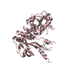
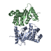
- PDB-5d65: X-RAY STRUCTURE OF MACROPHAGE INFLAMMATORY PROTEIN-1 ALPHA (CCL3)... -
+ Open data

- Basic information
| Entry | Database: PDB / ID: 5d65 | |||||||||
|---|---|---|---|---|---|---|---|---|---|---|
| Title | X-RAY STRUCTURE OF MACROPHAGE INFLAMMATORY PROTEIN-1 ALPHA (CCL3) WITH HEPARIN COMPLEX | |||||||||
 Components Components | C-C motif chemokine 3 | |||||||||
 Keywords Keywords | CYTOKINE / CC chemokine / CCL3 / OLIGOMER / SIGNALING PROTEIN / HEPARIN / GAG / COMPLEX | |||||||||
| Function / homology |  Function and homology information Function and homology informationlymphocyte chemotaxis / granulocyte chemotaxis / CCR1 chemokine receptor binding / positive regulation of natural killer cell chemotaxis / positive regulation of microglial cell migration / astrocyte cell migration / regulation of behavior / CCR5 chemokine receptor binding / eosinophil degranulation / CCR chemokine receptor binding ...lymphocyte chemotaxis / granulocyte chemotaxis / CCR1 chemokine receptor binding / positive regulation of natural killer cell chemotaxis / positive regulation of microglial cell migration / astrocyte cell migration / regulation of behavior / CCR5 chemokine receptor binding / eosinophil degranulation / CCR chemokine receptor binding / regulation of sensory perception of pain / signaling / positive regulation of microglial cell activation / cell activation / negative regulation of bone mineralization / T cell chemotaxis / eosinophil chemotaxis / positive regulation of calcium ion transport / response to cholesterol / chemokine activity / release of sequestered calcium ion into cytosol by sarcoplasmic reticulum / Chemokine receptors bind chemokines / phospholipase activator activity / chemoattractant activity / negative regulation of osteoclast differentiation / positive regulation of calcium ion import / Interleukin-10 signaling / exocytosis / macrophage chemotaxis / monocyte chemotaxis / host-mediated suppression of viral transcription / cellular response to interleukin-1 / neutrophil chemotaxis / cytoskeleton organization / positive regulation of calcium-mediated signaling / positive regulation of interleukin-1 beta production / chemokine-mediated signaling pathway / cell chemotaxis / calcium-mediated signaling / cellular response to type II interferon / cellular response to tumor necrosis factor / response to toxic substance / chemotaxis / intracellular calcium ion homeostasis / positive regulation of tumor necrosis factor production / positive regulation of inflammatory response / kinase activity / osteoblast differentiation / calcium ion transport / regulation of cell shape / positive regulation of neuron apoptotic process / MAPK cascade / cell-cell signaling / antimicrobial humoral immune response mediated by antimicrobial peptide / protein kinase activity / positive regulation of ERK1 and ERK2 cascade / positive regulation of phosphatidylinositol 3-kinase/protein kinase B signal transduction / positive regulation of cell migration / inflammatory response / negative regulation of gene expression / positive regulation of gene expression / : / extracellular region / identical protein binding / cytoplasm / cytosol Similarity search - Function | |||||||||
| Biological species |  Homo sapiens (human) Homo sapiens (human) | |||||||||
| Method |  X-RAY DIFFRACTION / SYNCHROTRON / SAD / Resolution: 3.1 Å X-RAY DIFFRACTION / SYNCHROTRON / SAD / Resolution: 3.1 Å | |||||||||
 Authors Authors | Liang, W.G. / Hwang, D.Y. / Zulueta, M.M. / Hung, S.C. / Tang, W. | |||||||||
| Funding support |  United States, 1items United States, 1items
| |||||||||
 Citation Citation | Journal: Proc.Natl.Acad.Sci.USA / Year: 2016 Title: Structural basis for oligomerization and glycosaminoglycan binding of CCL5 and CCL3. Authors: Liang, W.G. / Triandafillou, C.G. / Huang, T.Y. / Zulueta, M.M. / Banerjee, S. / Dinner, A.R. / Hung, S.C. / Tang, W.J. | |||||||||
| History |
|
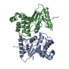
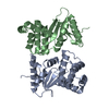
- Structure visualization
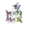
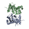
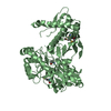
| Structure viewer | Molecule: MolmilJmol/JSmol |
|---|
- Downloads & links
-Download
| PDBx/mmCIF format | 5d65.cif.gz | 154.9 KB | Display | PDBx/mmCIF format |
|---|---|---|---|---|
| PDB format | pdb5d65.ent.gz | 124.8 KB | Display | PDB format |
| PDBx/mmJSON format | 5d65.json.gz | Tree view | PDBx/mmJSON format | |
| Others |  Other downloads Other downloads |
-Validation report
| Arichive directory | https://data.pdbj.org/pub/pdb/validation_reports/d6/5d65ftp://data.pdbj.org/pub/pdb/validation_reports/d6/5d65 | HTTPS FTP |
|---|
-Related structure data
| Related structure data | 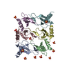 5cmdC 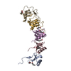 5corC 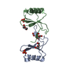 5coyC 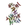 5dnfC 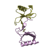 2x69S C: citing same article ( S: Starting model for refinement |
|---|---|
| Similar structure data |

-Links
 PDBj
PDBj


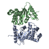
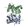
- Assembly
| Deposited unit | 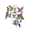
| ||||||||
|---|---|---|---|---|---|---|---|---|---|
| 1 |
| ||||||||
| Unit cell |
|
-Components
| #1: Protein | Mass: 7793.664 Da / Num. of mol.: 5 / Fragment: UNP residues 23-92 Source method: isolated from a genetically manipulated source Source: (gene. exp.) Homo sapiens (human) / Gene: CCL3, G0S19-1, MIP1A, SCYA3 / Production host:  #2: Polysaccharide | Source method: isolated from a genetically manipulated source #3: Sugar | ChemComp-BGC /   Type: D-saccharide, beta linking / Mass: 180.156 Da / Num. of mol.: 9 Type: D-saccharide, beta linking / Mass: 180.156 Da / Num. of mol.: 9Source method: isolated from a genetically manipulated source Formula: C6H12O6 #4: Chemical |   Mass: 35.453 Da / Num. of mol.: 2 / Source method: obtained synthetically / Formula: Cl Mass: 35.453 Da / Num. of mol.: 2 / Source method: obtained synthetically / Formula: Cl#5: Sugar | ChemComp-GLC / |   Type: D-saccharide, alpha linking / Mass: 180.156 Da / Num. of mol.: 1 Type: D-saccharide, alpha linking / Mass: 180.156 Da / Num. of mol.: 1Source method: isolated from a genetically manipulated source Formula: C6H12O6 Has protein modification | Y | |
|---|
-Experimental details
-Experiment
| Experiment | Method: X-RAY DIFFRACTION / Number of used crystals: 1 |
|---|
- Sample preparation
| Crystal | Density Matthews: 4.7 Å3/Da / Density % sol: 73.82 % |
|---|---|
| Crystal grow | Temperature: 291.15 K / Method: vapor diffusion, hanging drop / pH: 7.5 / Details: 0.1M Tris, pH 7.0; 1.8M (NH4)2SO4; / PH range: 7.0-7.8 |
-Data collection
| Diffraction | Mean temperature: 100 K |
|---|---|
| Diffraction source | Source: SYNCHROTRON / Site: APS / Beamline: 19-BM / Wavelength: 1.746 Å |
| Detector | Type: ADSC QUANTUM 210r / Detector: CCD / Date: Apr 11, 2014 |
| Radiation | Monochromator: Si(111) / Protocol: SINGLE WAVELENGTH / Monochromatic (M) / Laue (L): M / Scattering type: x-ray |
| Radiation wavelength | Wavelength: 1.746 Å / Relative weight: 1 |
| Reflection | Resolution: 3.095→34.22 Å / Num. obs: 14085 / % possible obs: 99.8 % / Observed criterion σ(I): 3 / Redundancy: 12.9 % / Rmerge(I) obs: 0.053 / Rsym value: 0.039 / Net I/σ(I): 48.9 |
| Reflection shell | Resolution: 3.1→3.13 Å / Redundancy: 8.1 % / Rmerge(I) obs: 0.678 / Mean I/σ(I) obs: 3 / % possible all: 100 |
- Processing
| Software |
| ||||||||||||||||||||||||||||||||||||||||||
|---|---|---|---|---|---|---|---|---|---|---|---|---|---|---|---|---|---|---|---|---|---|---|---|---|---|---|---|---|---|---|---|---|---|---|---|---|---|---|---|---|---|---|---|
| Refinement | Method to determine structure: SAD Starting model: 2X69 Resolution: 3.1→34.22 Å / SU ML: 0.33 / Cross valid method: FREE R-VALUE / σ(F): 0 / Phase error: 23.18 / Stereochemistry target values: MLHL
| ||||||||||||||||||||||||||||||||||||||||||
| Solvent computation | Shrinkage radii: 0.9 Å / VDW probe radii: 1.11 Å / Solvent model: FLAT BULK SOLVENT MODEL | ||||||||||||||||||||||||||||||||||||||||||
| Refinement step | Cycle: LAST / Resolution: 3.1→34.22 Å
| ||||||||||||||||||||||||||||||||||||||||||
| Refine LS restraints |
| ||||||||||||||||||||||||||||||||||||||||||
| LS refinement shell |
| ||||||||||||||||||||||||||||||||||||||||||
| Refinement TLS params. | Method: refined / Origin x: 22.7769 Å / Origin y: 73.9483 Å / Origin z: 46.6755 Å
| ||||||||||||||||||||||||||||||||||||||||||
| Refinement TLS group | Selection details: ALL |
