[English] 日本語
Yorodumi
- PDB-5d3q: Dynamin 1 GTPase-BSE fusion dimer complexed with GDP -

+
Open data


ID or keywords:

Loading...

-
Basic information

Entry
Database: PDB / ID: 5d3q
TitleDynamin 1 GTPase-BSE fusion dimer complexed with GDP
ComponentsDynamin-1,Dynamin-1
KeywordsHYDROLASE / Fusion protein / GTPase / Endocytosis
Function / homology
Function and homology information


clathrin coat assembly involved in endocytosis / vesicle scission / synaptic vesicle budding from presynaptic endocytic zone membrane / presynaptic endocytic zone membrane / dynamin GTPase / chromaffin granule / regulation of vesicle size / Toll Like Receptor 4 (TLR4) Cascade / Retrograde neurotrophin signalling / Formation of annular gap junctions ...clathrin coat assembly involved in endocytosis / vesicle scission / synaptic vesicle budding from presynaptic endocytic zone membrane / presynaptic endocytic zone membrane / dynamin GTPase / chromaffin granule / regulation of vesicle size / Toll Like Receptor 4 (TLR4) Cascade / Retrograde neurotrophin signalling / Formation of annular gap junctions / endosome organization / Gap junction degradation / photoreceptor ribbon synapse / membrane coat / Recycling pathway of L1 / phosphatidylinositol-3,4,5-trisphosphate binding / endocytic vesicle / EPH-ephrin mediated repulsion of cells / clathrin-coated pit / phosphatidylinositol-4,5-bisphosphate binding / MHC class II antigen presentation / photoreceptor inner segment / receptor-mediated endocytosis / cell projection / modulation of chemical synaptic transmission / protein homooligomerization / receptor internalization / endocytosis / : / GDP binding / presynapse / Clathrin-mediated endocytosis / microtubule binding / protein homotetramerization / microtubule / GTPase activity / glutamatergic synapse / synapse / GTP binding / protein kinase binding / protein homodimerization activity / RNA binding / extracellular exosome / identical protein binding / plasma membrane / cytoplasm
Similarity search - Function
Dynamin GTPase effector / Dynamin GTPase effector domain / Dynamin GTPase effector domain / Dynamin, GTPase region, conserved site / Dynamin-type guanine nucleotide-binding (G) domain signature. / Dynamin stalk domain / Dynamin central region / GTPase effector domain / GED domain profile. / Dynamin, GTPase domain ...Dynamin GTPase effector / Dynamin GTPase effector domain / Dynamin GTPase effector domain / Dynamin, GTPase region, conserved site / Dynamin-type guanine nucleotide-binding (G) domain signature. / Dynamin stalk domain / Dynamin central region / GTPase effector domain / GED domain profile. / Dynamin, GTPase domain / Dynamin, GTPase / Dynamin / Dynamin-type guanine nucleotide-binding (G) domain / Dynamin-type guanine nucleotide-binding (G) domain profile. / Dynamin, N-terminal / Dynamin family / PH domain / PH domain profile. / Pleckstrin homology domain. / Pleckstrin homology domain / PH-like domain superfamily / P-loop containing nucleotide triphosphate hydrolases / Rossmann fold / P-loop containing nucleoside triphosphate hydrolase / 3-Layer(aba) Sandwich / Alpha Beta
Similarity search - Domain/homology
GUANOSINE-5'-DIPHOSPHATE / Dynamin-1
Similarity search - Component
Biological speciesHomo sapiens (human)
MethodX-RAY DIFFRACTION / SYNCHROTRON / MOLECULAR REPLACEMENT / molecular replacement / Resolution: 1.7 Å
AuthorsAnand, R. / Eschenburg, S. / Reubold, T.F.
CitationJournal: Biochem.Biophys.Res.Commun. / Year: 2016
Title: Crystal structure of the GTPase domain and the bundle signalling element of dynamin in the GDP state.
Authors: Anand, R. / Eschenburg, S. / Reubold, T.F.
History
DepositionAug 6, 2015Deposition site: RCSB / Processing site: PDBE
Revision 1.0Dec 2, 2015Provider: repository / Type: Initial release
Revision 1.1Sep 14, 2016Group: Database references
Revision 1.2Mar 22, 2017Group: Data collection
Revision 1.3Jan 10, 2024Group: Data collection / Database references / Refinement description
Category: chem_comp_atom / chem_comp_bond ...chem_comp_atom / chem_comp_bond / database_2 / pdbx_initial_refinement_model
Item: _database_2.pdbx_DOI / _database_2.pdbx_database_accession

-
Structure visualization

Structure viewerMolecule:
MolmilJmol/JSmol

Downloads & links

-
Assembly

Deposited unit
A: Dynamin-1,Dynamin-1
B: Dynamin-1,Dynamin-1
hetero molecules


Theoretical massNumber of molelcules
Total (without water)78,0656
Polymers77,0542
Non-polymers1,0114
Water10,971609
1
A: Dynamin-1,Dynamin-1
hetero molecules


Theoretical massNumber of molelcules
Total (without water)39,0323
Polymers38,5271
Non-polymers5052
Water181
TypeNameSymmetry operationNumber
identity operation1_555x,y,z1
2
B: Dynamin-1,Dynamin-1
hetero molecules


Theoretical massNumber of molelcules
Total (without water)39,0323
Polymers38,5271
Non-polymers5052
Water181
TypeNameSymmetry operationNumber
identity operation1_555x,y,z1
Unit cell
Length a, b, c (Å)44.220, 49.960, 82.520
Angle α, β, γ (deg.)99.40, 87.18, 105.18
Int Tables number1
Space group name H-MP1

-
Components

#1: Protein Dynamin-1,Dynamin-1


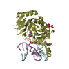
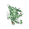
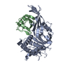
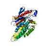
Mass: 38527.035 Da / Num. of mol.: 2
Fragment: UNP residues 5-320, 726-746,UNP residues 5-320, 726-746
Source method: isolated from a genetically manipulated source
Details: The sequence contains residues 5-320 and 726-746 of human dynamin 1. The very N-terminus contains a glycine residue from the TEV cleavage step. The two dynamin fragments are connected by a ...Details: The sequence contains residues 5-320 and 726-746 of human dynamin 1. The very N-terminus contains a glycine residue from the TEV cleavage step. The two dynamin fragments are connected by a linker sequence of eight residues (KHGTDSRV).
Source: (gene. exp.) Homo sapiens (human) / Gene: DNM1, DNM / Production host: Escherichia coli BL21(DE3) (bacteria) / Variant (production host): Rosetta / References: UniProt: Q05193, dynamin GTPase
#2: Chemical ChemComp-GDP / GUANOSINE-5'-DIPHOSPHATE


Type: RNA linking / Mass: 443.201 Da / Num. of mol.: 2 / Source method: obtained synthetically / Formula: C10H15N5O11P2 / Comment: GDP, energy-carrying molecule*YM
#3: Chemical ChemComp-EDO / 1,2-ETHANEDIOL / ETHYLENE GLYCOL


Mass: 62.068 Da / Num. of mol.: 2 / Source method: obtained synthetically / Formula: C2H6O2
#4: Water ChemComp-HOH / water


Mass: 18.015 Da / Num. of mol.: 609 / Source method: isolated from a natural source / Formula: H2O

-
Experimental details

-
Experiment

ExperimentMethod: X-RAY DIFFRACTION / Number of used crystals: 1

-
Sample preparation

CrystalDensity Matthews: 2.25 Å3/Da / Density % sol: 45.4 %
Crystal growTemperature: 277 K / Method: vapor diffusion, hanging drop / pH: 8
Details: Before crystallization set-ups, GG1 at a concentration of 10 mg/ml was supplemented with 2 mM GDP and 2 mM MgCl2 and incubated for 1 h on ice. GG1GDP was crystallized by hanging drop vapour ...Details: Before crystallization set-ups, GG1 at a concentration of 10 mg/ml was supplemented with 2 mM GDP and 2 mM MgCl2 and incubated for 1 h on ice. GG1GDP was crystallized by hanging drop vapour diffusion mixing 1.5 ul protein solution with 1.5 ul reservoir solution containing 0.1 M Tris (pH 8.0), 26 % PEG 3350 and 0.2 M NaSCN. The drops were equilibrated against 700 ul of reservoir.
PH range: 8

-
Data collection

DiffractionMean temperature: 100 K
Diffraction sourceSource: SYNCHROTRON / Site: PETRA III, DESY / Beamline: P11 / Wavelength: 1.033 Å
DetectorType: PSI PILATUS 6M / Detector: PIXEL / Date: Mar 12, 2013
RadiationProtocol: SINGLE WAVELENGTH / Monochromatic (M) / Laue (L): M / Scattering type: x-ray
Radiation wavelengthWavelength: 1.033 Å / Relative weight: 1
ReflectionResolution: 1.7→47.626 Å / Num. obs: 72213 / % possible obs: 97.6 % / Redundancy: 4.63 % / Rmerge(I) obs: 0.118 / Rsym value: 0.108 / Net I/σ(I): 10.75
Reflection shellResolution: 1.7→1.8 Å / Redundancy: 3.85 % / Rmerge(I) obs: 0.245 / Mean I/σ(I) obs: 6.7 / % possible all: 96.1

-
Phasing

PhasingMethod: molecular replacement

-
Processing

Software
NameVersionClassification
PHENIX(1.10_2155: ???)refinement
XSCALEdata scaling
PHASERphasing
PDB_EXTRACT3.15data extraction
XDSdata reduction
Cootmodel building
RefinementMethod to determine structure: MOLECULAR REPLACEMENT
Starting model: 2x2e
Resolution: 1.7→47.626 Å / SU ML: 0.18 / Cross valid method: FREE R-VALUE / σ(F): 2 / Phase error: 21.7 / Stereochemistry target values: ML
RfactorNum. reflection% reflectionSelection details
Rfree0.2055 3611 5 %Random selection
Rwork0.1727 ---
obs0.1744 72207 97.6 %-
Solvent computationShrinkage radii: 0.9 Å / VDW probe radii: 1.11 Å / Solvent model: FLAT BULK SOLVENT MODEL
Refinement stepCycle: LAST / Resolution: 1.7→47.626 Å
ProteinNucleic acidLigandSolventTotal
Num. atoms5134 0 64 609 5807
Refine LS restraints
Refine-IDTypeDev idealNumber
X-RAY DIFFRACTIONf_bond_d0.0155347
X-RAY DIFFRACTIONf_angle_d1.47259
X-RAY DIFFRACTIONf_dihedral_angle_d13.6583346
X-RAY DIFFRACTIONf_chiral_restr0.082844
X-RAY DIFFRACTIONf_plane_restr0.008945
LS refinement shell
Resolution (Å)Rfactor RfreeNum. reflection RfreeRfactor RworkNum. reflection RworkRefine-ID% reflection obs (%)
1.7-1.72240.26571390.20692642X-RAY DIFFRACTION96
1.7224-1.7460.25881340.21052538X-RAY DIFFRACTION96
1.746-1.77090.24681370.20122603X-RAY DIFFRACTION96
1.7709-1.79730.24711360.19642599X-RAY DIFFRACTION96
1.7973-1.82540.23251380.19472616X-RAY DIFFRACTION96
1.8254-1.85540.25671380.18382620X-RAY DIFFRACTION96
1.8554-1.88740.21591340.1812552X-RAY DIFFRACTION97
1.8874-1.92170.23181420.18192690X-RAY DIFFRACTION97
1.9217-1.95860.18791330.17952538X-RAY DIFFRACTION96
1.9586-1.99860.23851400.17812657X-RAY DIFFRACTION97
1.9986-2.04210.23241360.18132579X-RAY DIFFRACTION97
2.0421-2.08960.2241390.18262644X-RAY DIFFRACTION97
2.0896-2.14180.21851370.17552596X-RAY DIFFRACTION97
2.1418-2.19970.20431400.17652653X-RAY DIFFRACTION97
2.1997-2.26450.22291370.17782608X-RAY DIFFRACTION98
2.2645-2.33760.21891400.17412661X-RAY DIFFRACTION97
2.3376-2.42110.21761380.17812620X-RAY DIFFRACTION97
2.4211-2.5180.22271390.17222639X-RAY DIFFRACTION98
2.518-2.63260.23321420.1742709X-RAY DIFFRACTION99
2.6326-2.77140.21291420.17712691X-RAY DIFFRACTION100
2.7714-2.9450.22091400.17822673X-RAY DIFFRACTION100
2.945-3.17240.22151430.18112702X-RAY DIFFRACTION100
3.1724-3.49150.18831400.17392658X-RAY DIFFRACTION100
3.4915-3.99650.16841430.15732720X-RAY DIFFRACTION100
3.9965-5.03430.16961410.14672683X-RAY DIFFRACTION100
5.0343-47.64470.17931430.16822705X-RAY DIFFRACTION100

+
About Yorodumi

-
News

-
Feb 9, 2022. New format data for meta-information of EMDB entries

New format data for meta-information of EMDB entries

  • Version 3 of the EMDB header file is now the official format.
  • The previous official version 1.9 will be removed from the archive.

Related info.:EMDB header

External links:wwPDB to switch to version 3 of the EMDB data model

-
Aug 12, 2020. Covid-19 info

Covid-19 info

URL: https://pdbjlvh1.pdbj.org/emnavi/covid19.php

New page: Covid-19 featured information page in EM Navigator.

Related info.:Covid-19 info / Mar 5, 2020. Novel coronavirus structure data

+
Mar 5, 2020. Novel coronavirus structure data

Novel coronavirus structure data

Related info.:Yorodumi Speices / Aug 12, 2020. Covid-19 info

External links:COVID-19 featured content - PDBj / Molecule of the Month (242):Coronavirus Proteases

+
Jan 31, 2019. EMDB accession codes are about to change! (news from PDBe EMDB page)

EMDB accession codes are about to change! (news from PDBe EMDB page)

  • The allocation of 4 digits for EMDB accession codes will soon come to an end. Whilst these codes will remain in use, new EMDB accession codes will include an additional digit and will expand incrementally as the available range of codes is exhausted. The current 4-digit format prefixed with “EMD-” (i.e. EMD-XXXX) will advance to a 5-digit format (i.e. EMD-XXXXX), and so on. It is currently estimated that the 4-digit codes will be depleted around Spring 2019, at which point the 5-digit format will come into force.
  • The EM Navigator/Yorodumi systems omit the EMD- prefix.

Related info.:Q: What is EMD? / ID/Accession-code notation in Yorodumi/EM Navigator

External links:EMDB Accession Codes are Changing Soon! / Contact to PDBj

+
Jul 12, 2017. Major update of PDB

Major update of PDB

  • wwPDB released updated PDB data conforming to the new PDBx/mmCIF dictionary.
  • This is a major update changing the version number from 4 to 5, and with Remediation, in which all the entries are updated.
  • In this update, many items about electron microscopy experimental information are reorganized (e.g. em_software).
  • Now, EM Navigator and Yorodumi are based on the updated data.

External links:wwPDB Remediation / Enriched Model Files Conforming to OneDep Data Standards Now Available in the PDB FTP Archive

-
Yorodumi

Thousand views of thousand structures

  • Yorodumi is a browser for structure data from EMDB, PDB, SASBDB, etc.
  • This page is also the successor to EM Navigator detail page, and also detail information page/front-end page for Omokage search.
  • The word "yorodu" (or yorozu) is an old Japanese word meaning "ten thousand". "mi" (miru) is to see.

Related info.:EMDB / PDB / SASBDB / Comparison of 3 databanks / Yorodumi Search / Aug 31, 2016. New EM Navigator & Yorodumi / Yorodumi Papers / Jmol/JSmol / Function and homology information / Changes in new EM Navigator and Yorodumi

Read more