[English] 日本語
Yorodumi
- PDB-5cyv: Crystal structure of CouR from Rhodococcus jostii RHA1 bound to p... -

+
Open data


ID or keywords:

Loading...

-
Basic information

Entry
Database: PDB / ID: 5cyv
Title
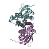
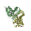
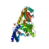
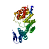
Crystal structure of CouR from Rhodococcus jostii RHA1 bound to p-coumaroyl-CoA
ComponentsTranscriptional regulator
KeywordsTranscriptional regulator / TRANSCRIPTION REGULATOR / p-hydroxycinnamate metabolism / MCSG / PF04017 / PSI-BIOLOGY / MARR / STRUCTURAL GENOMICS / PROTEIN STRUCTURE INITIATIVE / MIDWEST CENTER FOR STRUCTURAL GENOMICS
Function / homology
Function and homology information


DNA-binding transcription factor activity / metal ion binding
Similarity search - Function
Transcriptional regulator MarR-type, conserved site / MarR-type HTH domain signature. / MarR family / : / MarR family / MarR-type HTH domain profile. / helix_turn_helix multiple antibiotic resistance protein / MarR-type HTH domain / Winged helix-like DNA-binding domain superfamily/Winged helix DNA-binding domain / Arc Repressor Mutant, subunit A ...Transcriptional regulator MarR-type, conserved site / MarR-type HTH domain signature. / MarR family / : / MarR family / MarR-type HTH domain profile. / helix_turn_helix multiple antibiotic resistance protein / MarR-type HTH domain / Winged helix-like DNA-binding domain superfamily/Winged helix DNA-binding domain / Arc Repressor Mutant, subunit A / Winged helix DNA-binding domain superfamily / Winged helix-like DNA-binding domain superfamily / Orthogonal Bundle / Mainly Alpha
Similarity search - Domain/homology
ACETATE ION / p-coumaroyl-CoA / Transcriptional regulator
Similarity search - Component
Biological speciesRhodococcus jostii (bacteria)
MethodX-RAY DIFFRACTION / MOLECULAR REPLACEMENT / Resolution: 1.52 Å
AuthorsStogios, P.J. / Xu, X. / Dong, A. / Savchenko, A. / Joachimiak, A. / Midwest Center for Structural Genomics (MCSG)
Funding support United States, 1items
OrganizationGrant numberCountry
Department of Energy (DOE, United States)DE-AC02-06CH11357 United States
CitationJournal: Nucleic Acids Res. / Year: 2016
Title: The activity of CouR, a MarR family transcriptional regulator, is modulated through a novel molecular mechanism.
Authors: Otani, H. / Stogios, P.J. / Xu, X. / Nocek, B. / Li, S.N. / Savchenko, A. / Eltis, L.D.
History
DepositionJul 30, 2015Deposition site: RCSB / Processing site: RCSB
Revision 1.0Aug 12, 2015Provider: repository / Type: Initial release
Revision 1.1Mar 23, 2016Group: Database references
Revision 1.2Sep 20, 2017Group: Author supporting evidence / Database references / Derived calculations
Category: citation / pdbx_audit_support / pdbx_struct_oper_list
Item: _citation.journal_id_CSD / _pdbx_audit_support.funding_organization / _pdbx_struct_oper_list.symmetry_operation
Revision 1.3Nov 1, 2017Group: Author supporting evidence / Category: pdbx_struct_assembly_auth_evidence
Revision 1.4Dec 4, 2019Group: Author supporting evidence / Category: pdbx_audit_support / Item: _pdbx_audit_support.funding_organization
Revision 1.5Sep 27, 2023Group: Data collection / Database references ...Data collection / Database references / Derived calculations / Refinement description
Category: chem_comp_atom / chem_comp_bond ...chem_comp_atom / chem_comp_bond / database_2 / pdbx_initial_refinement_model / pdbx_struct_conn_angle / struct_conn
Item: _database_2.pdbx_DOI / _database_2.pdbx_database_accession ..._database_2.pdbx_DOI / _database_2.pdbx_database_accession / _pdbx_struct_conn_angle.ptnr1_auth_asym_id / _pdbx_struct_conn_angle.ptnr1_auth_comp_id / _pdbx_struct_conn_angle.ptnr1_auth_seq_id / _pdbx_struct_conn_angle.ptnr1_label_asym_id / _pdbx_struct_conn_angle.ptnr1_label_atom_id / _pdbx_struct_conn_angle.ptnr1_label_comp_id / _pdbx_struct_conn_angle.ptnr1_symmetry / _pdbx_struct_conn_angle.ptnr3_auth_asym_id / _pdbx_struct_conn_angle.ptnr3_auth_comp_id / _pdbx_struct_conn_angle.ptnr3_auth_seq_id / _pdbx_struct_conn_angle.ptnr3_label_asym_id / _pdbx_struct_conn_angle.ptnr3_label_atom_id / _pdbx_struct_conn_angle.ptnr3_label_comp_id / _pdbx_struct_conn_angle.ptnr3_symmetry / _pdbx_struct_conn_angle.value / _struct_conn.pdbx_dist_value / _struct_conn.ptnr1_auth_seq_id / _struct_conn.ptnr1_label_asym_id / _struct_conn.ptnr2_auth_asym_id / _struct_conn.ptnr2_auth_comp_id / _struct_conn.ptnr2_auth_seq_id / _struct_conn.ptnr2_label_asym_id / _struct_conn.ptnr2_label_atom_id / _struct_conn.ptnr2_label_comp_id / _struct_conn.ptnr2_symmetry
Revision 1.6Mar 13, 2024Group: Source and taxonomy / Category: entity_src_gen

-
Structure visualization

Structure viewerMolecule:
MolmilJmol/JSmol

Downloads & links

-
Assembly

Deposited unit
A: Transcriptional regulator
B: Transcriptional regulator
hetero molecules


Theoretical massNumber of molelcules
Total (without water)33,72411
Polymers31,6582
Non-polymers2,0669
Water8,143452
1


  • Idetical with deposited unit
  • defined by author&software
  • Evidence: gel filtration, light scattering
TypeNameSymmetry operationNumber
identity operation1_555x,y,z1
Buried area5810 Å2
ΔGint-82 kcal/mol
Surface area14490 Å2
MethodPISA
Unit cell
Length a, b, c (Å)64.145, 134.385, 73.336
Angle α, β, γ (deg.)90.00, 90.00, 90.00
Int Tables number20
Space group name H-MC2221
Components on special symmetry positions
IDModelComponents
11B-500-

HOH

21B-514-

HOH

31B-537-

HOH

DetailsDimer according to SEC-MALS

-
Components

-
Protein , 1 types, 2 molecules AB

#1: Protein Transcriptional regulator


Mass: 15828.976 Da / Num. of mol.: 2
Source method: isolated from a genetically manipulated source
Source: (gene. exp.) Rhodococcus jostii (strain RHA1) (bacteria)
Strain: RHA1 / Gene: RHA1_ro05125 / Plasmid: p15TV-LIC / Production host: Escherichia coli (E. coli) / Strain (production host): BL21(DE3) / References: UniProt: Q0S6D0

-
Non-polymers , 5 types, 461 molecules

#2: Chemical ChemComp-MG / MAGNESIUM ION


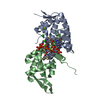
Mass: 24.305 Da / Num. of mol.: 3 / Source method: obtained synthetically / Formula: Mg
#3: Chemical ChemComp-ACT / ACETATE ION


Mass: 59.044 Da / Num. of mol.: 1 / Source method: obtained synthetically / Formula: C2H3O2
#4: Chemical ChemComp-CL / CHLORIDE ION


Mass: 35.453 Da / Num. of mol.: 3 / Source method: obtained synthetically / Formula: Cl
#5: Chemical ChemComp-WCA / p-coumaroyl-CoA


Mass: 913.677 Da / Num. of mol.: 2
Source method: isolated from a genetically manipulated source
Formula: C30H42N7O18P3S
#6: Water ChemComp-HOH / water


Mass: 18.015 Da / Num. of mol.: 452 / Source method: isolated from a natural source / Formula: H2O

-
Experimental details

-
Experiment

ExperimentMethod: X-RAY DIFFRACTION

-
Sample preparation

CrystalDensity Matthews: 2.5 Å3/Da / Density % sol: 50.7 %
Crystal growTemperature: 298 K / Method: vapor diffusion, hanging drop / pH: 6.5
Details: 80 microL of native protein at 21 mg/ml were preincubated with 20 microL of 25 mM p-hydroxycinnamoyl-CoA. Reservoir = 0.2 M magnesium acetate, 0.1 M sodium cacodylate pH 6.5, 4% (w/v) 2- ...Details: 80 microL of native protein at 21 mg/ml were preincubated with 20 microL of 25 mM p-hydroxycinnamoyl-CoA. Reservoir = 0.2 M magnesium acetate, 0.1 M sodium cacodylate pH 6.5, 4% (w/v) 2-methyl-2,4-pentanediol and 26% (w/v) PEG 8K

-
Data collection

DiffractionMean temperature: 100 K
Diffraction sourceSource: ROTATING ANODE / Type: RIGAKU MICROMAX-007 HF / Wavelength: 1.5418 Å
DetectorType: RIGAKU RAXIS IV++ / Detector: IMAGE PLATE / Date: Mar 22, 2014
RadiationMonochromator: graphite / Protocol: SINGLE WAVELENGTH / Monochromatic (M) / Laue (L): M / Scattering type: x-ray
Radiation wavelengthWavelength: 1.5418 Å / Relative weight: 1
ReflectionResolution: 1.51→25 Å / Num. obs: 48946 / % possible obs: 97.8 % / Redundancy: 6.7 % / Rmerge(I) obs: 0.033 / Net I/σ(I): 67
Reflection shellResolution: 1.51→1.54 Å / Redundancy: 4.4 % / Rmerge(I) obs: 0.47 / Mean I/σ(I) obs: 3.79 / % possible all: 83.6

-
Processing

Software
NameVersionClassification
HKL-3000data reduction
HKL-3000data scaling
PHENIXphasing
PHENIX1.9_1692refinement
Cootmodel building
RefinementMethod to determine structure: MOLECULAR REPLACEMENT
Starting model: 3FM5
Resolution: 1.52→24.789 Å / SU ML: 0.18 / Cross valid method: FREE R-VALUE / σ(F): 1.34 / Phase error: 21.46 / Stereochemistry target values: ML
RfactorNum. reflection% reflectionSelection details
Rfree0.2017 3774 4.09 %Random selection
Rwork0.1606 ---
obs0.1623 48184 97.81 %-
Solvent computationShrinkage radii: 0.9 Å / VDW probe radii: 1.11 Å / Solvent model: FLAT BULK SOLVENT MODEL
Refinement stepCycle: LAST / Resolution: 1.52→24.789 Å
ProteinNucleic acidLigandSolventTotal
Num. atoms2244 0 10 452 2706
Refine LS restraints
Refine-IDTypeDev idealNumber
X-RAY DIFFRACTIONf_bond_d0.0182344
X-RAY DIFFRACTIONf_angle_d1.853173
X-RAY DIFFRACTIONf_dihedral_angle_d22.358977
X-RAY DIFFRACTIONf_chiral_restr0.084355
X-RAY DIFFRACTIONf_plane_restr0.009411
LS refinement shellResolution: 1.52→1.5393 Å
RfactorNum. reflection% reflection
Rfree0.2747 127 -
Rwork0.2673 2788 -
obs--83 %

+
About Yorodumi

-
News

-
Feb 9, 2022. New format data for meta-information of EMDB entries

New format data for meta-information of EMDB entries

  • Version 3 of the EMDB header file is now the official format.
  • The previous official version 1.9 will be removed from the archive.

Related info.:EMDB header

External links:wwPDB to switch to version 3 of the EMDB data model

-
Aug 12, 2020. Covid-19 info

Covid-19 info

URL: https://pdbjlvh1.pdbj.org/emnavi/covid19.php

New page: Covid-19 featured information page in EM Navigator.

Related info.:Covid-19 info / Mar 5, 2020. Novel coronavirus structure data

+
Mar 5, 2020. Novel coronavirus structure data

Novel coronavirus structure data

Related info.:Yorodumi Speices / Aug 12, 2020. Covid-19 info

External links:COVID-19 featured content - PDBj / Molecule of the Month (242):Coronavirus Proteases

+
Jan 31, 2019. EMDB accession codes are about to change! (news from PDBe EMDB page)

EMDB accession codes are about to change! (news from PDBe EMDB page)

  • The allocation of 4 digits for EMDB accession codes will soon come to an end. Whilst these codes will remain in use, new EMDB accession codes will include an additional digit and will expand incrementally as the available range of codes is exhausted. The current 4-digit format prefixed with “EMD-” (i.e. EMD-XXXX) will advance to a 5-digit format (i.e. EMD-XXXXX), and so on. It is currently estimated that the 4-digit codes will be depleted around Spring 2019, at which point the 5-digit format will come into force.
  • The EM Navigator/Yorodumi systems omit the EMD- prefix.

Related info.:Q: What is EMD? / ID/Accession-code notation in Yorodumi/EM Navigator

External links:EMDB Accession Codes are Changing Soon! / Contact to PDBj

+
Jul 12, 2017. Major update of PDB

Major update of PDB

  • wwPDB released updated PDB data conforming to the new PDBx/mmCIF dictionary.
  • This is a major update changing the version number from 4 to 5, and with Remediation, in which all the entries are updated.
  • In this update, many items about electron microscopy experimental information are reorganized (e.g. em_software).
  • Now, EM Navigator and Yorodumi are based on the updated data.

External links:wwPDB Remediation / Enriched Model Files Conforming to OneDep Data Standards Now Available in the PDB FTP Archive

-
Yorodumi

Thousand views of thousand structures

  • Yorodumi is a browser for structure data from EMDB, PDB, SASBDB, etc.
  • This page is also the successor to EM Navigator detail page, and also detail information page/front-end page for Omokage search.
  • The word "yorodu" (or yorozu) is an old Japanese word meaning "ten thousand". "mi" (miru) is to see.

Related info.:EMDB / PDB / SASBDB / Comparison of 3 databanks / Yorodumi Search / Aug 31, 2016. New EM Navigator & Yorodumi / Yorodumi Papers / Jmol/JSmol / Function and homology information / Changes in new EM Navigator and Yorodumi

Read more