 Movie
Movie Controller

[English] 日本語
 Yorodumi
- PDB-5cuy: Crystal structure of Trypanosoma brucei Vacuolar Soluble Pyrophos... -
+ Open data

- Basic information
| Entry | Database: PDB / ID: 5cuy | ||||||
|---|---|---|---|---|---|---|---|
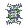
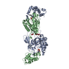
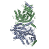
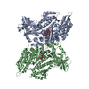
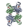
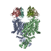
| Title | Crystal structure of Trypanosoma brucei Vacuolar Soluble Pyrophosphatases in apo form | ||||||
 Components Components | Acidocalcisomal pyrophosphatase | ||||||
 Keywords Keywords | METAL BINDING PROTEIN / substrate binding / acidocalcisomal pyrophosphatase | ||||||
| Function / homology |  Function and homology information Function and homology informationacidocalcisome / pyrophosphatase activity / inorganic diphosphatase / inorganic diphosphate phosphatase activity / phosphate-containing compound metabolic process / calcium ion binding / magnesium ion binding / zinc ion binding / cytoplasm Similarity search - Function | ||||||
| Biological species |  | ||||||
| Method |  X-RAY DIFFRACTION / SYNCHROTRON / MOLECULAR REPLACEMENT / Resolution: 2.5 Å X-RAY DIFFRACTION / SYNCHROTRON / MOLECULAR REPLACEMENT / Resolution: 2.5 Å | ||||||
 Authors Authors | Yang, Y.Y. / Ko, T.P. / Liu, W.D. / Zheng, Y.Y. / Chen, C.C. / Guo, R.T. | ||||||
 Citation Citation | Journal: Acs Chem.Biol. / Year: 2016 Title: Crystal structure of Trypanosoma cruzi protein in complex with ligand Authors: Yang, Y.Y. / Ko, T.P. / Zheng, Y.Y. / Liu, W.D. / Chen, C.C. / Guo, R.T. | ||||||
| History |
|
- Structure visualization
| Structure viewer | Molecule: MolmilJmol/JSmol |
|---|
- Downloads & links
-Download
| PDBx/mmCIF format | 5cuy.cif.gz | 336.6 KB | Display | PDBx/mmCIF format |
|---|---|---|---|---|
| PDB format | pdb5cuy.ent.gz | 273.4 KB | Display | PDB format |
| PDBx/mmJSON format | 5cuy.json.gz | Tree view | PDBx/mmJSON format | |
| Others |  Other downloads Other downloads |
-Validation report
| Arichive directory | https://data.pdbj.org/pub/pdb/validation_reports/cu/5cuyftp://data.pdbj.org/pub/pdb/validation_reports/cu/5cuy | HTTPS FTP |
|---|
-Related structure data
| Related structure data |  5cuuC  5cuvSC  5cuxC C: citing same article ( S: Starting model for refinement |
|---|---|
| Similar structure data |


-Links
 PDBj
PDBj- Assembly
| Deposited unit | 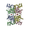
| ||||||||
|---|---|---|---|---|---|---|---|---|---|
| 1 |
| ||||||||
| Unit cell |
|
-Components
| #1: Protein | Mass: 47403.629 Da / Num. of mol.: 4 Source method: isolated from a genetically manipulated source Source: (gene. exp.) Strain: 927/4 GUTat10.1 / Gene: Tb11.02.4930 / Plasmid: pET-32 Xa/LIC / Production host:  #2: Chemical | ChemComp-CIT /   Mass: 192.124 Da / Num. of mol.: 4 / Source method: obtained synthetically / Formula: C6H8O7 Mass: 192.124 Da / Num. of mol.: 4 / Source method: obtained synthetically / Formula: C6H8O7#3: Chemical | ChemComp-MG /   Mass: 24.305 Da / Num. of mol.: 4 / Source method: obtained synthetically / Formula: Mg Mass: 24.305 Da / Num. of mol.: 4 / Source method: obtained synthetically / Formula: Mg#4: Water | ChemComp-HOH / |  Mass: 18.015 Da / Num. of mol.: 686 / Source method: isolated from a natural source / Formula: H2O Mass: 18.015 Da / Num. of mol.: 686 / Source method: isolated from a natural source / Formula: H2O |
|---|
-Experimental details
-Experiment
| Experiment | Method: X-RAY DIFFRACTION / Number of used crystals: 1 |
|---|
- Sample preparation
| Crystal | Density Matthews: 2.63 Å3/Da / Density % sol: 53.15 % |
|---|---|
| Crystal grow | Temperature: 298 K / Method: vapor diffusion, sitting drop / pH: 8.3 / Details: potassium citrate tribasic,PEG 3350 |
-Data collection
| Diffraction | Mean temperature: 100 K | |||||||||||||||||||||||||||||||||||||||||||||||||||||||||||||||||||||||||||||||||||||||||||||||||||
|---|---|---|---|---|---|---|---|---|---|---|---|---|---|---|---|---|---|---|---|---|---|---|---|---|---|---|---|---|---|---|---|---|---|---|---|---|---|---|---|---|---|---|---|---|---|---|---|---|---|---|---|---|---|---|---|---|---|---|---|---|---|---|---|---|---|---|---|---|---|---|---|---|---|---|---|---|---|---|---|---|---|---|---|---|---|---|---|---|---|---|---|---|---|---|---|---|---|---|---|---|
| Diffraction source | Source: SYNCHROTRON / Site: NSRRC  / Beamline: BL13C1 / Wavelength: 0.97622 Å / Beamline: BL13C1 / Wavelength: 0.97622 Å | |||||||||||||||||||||||||||||||||||||||||||||||||||||||||||||||||||||||||||||||||||||||||||||||||||
| Detector | Type: RAYONIX MX300HE / Detector: CCD / Date: Nov 22, 2014 | |||||||||||||||||||||||||||||||||||||||||||||||||||||||||||||||||||||||||||||||||||||||||||||||||||
| Radiation | Monochromator: GRAPHITE / Protocol: SINGLE WAVELENGTH / Monochromatic (M) / Laue (L): M / Scattering type: x-ray | |||||||||||||||||||||||||||||||||||||||||||||||||||||||||||||||||||||||||||||||||||||||||||||||||||
| Radiation wavelength | Wavelength: 0.97622 Å / Relative weight: 1 | |||||||||||||||||||||||||||||||||||||||||||||||||||||||||||||||||||||||||||||||||||||||||||||||||||
| Reflection | Resolution: 2.5→25 Å / Num. obs: 64763 / % possible obs: 98.8 % / Redundancy: 3.3 % / Rmerge(I) obs: 0.09 / Rpim(I) all: 0.058 / Rrim(I) all: 0.108 / Χ2: 1.185 / Net I/av σ(I): 15.394 / Net I/σ(I): 9.6 / Num. measured all: 216422 | |||||||||||||||||||||||||||||||||||||||||||||||||||||||||||||||||||||||||||||||||||||||||||||||||||
| Reflection shell | Diffraction-ID: 1 / Rejects: _
|
- Processing
| Software |
| ||||||||||||||||||||||||
|---|---|---|---|---|---|---|---|---|---|---|---|---|---|---|---|---|---|---|---|---|---|---|---|---|---|
| Refinement | Method to determine structure: MOLECULAR REPLACEMENT Starting model: 5CUV Resolution: 2.5→25 Å / σ(F): 0
| ||||||||||||||||||||||||
| Solvent computation | Bsol: 28.9844 Å2 | ||||||||||||||||||||||||
| Displacement parameters | Biso max: 121.04 Å2 / Biso mean: 50.4658 Å2 / Biso min: 16.13 Å2
| ||||||||||||||||||||||||
| Refinement step | Cycle: final / Resolution: 2.5→25 Å
| ||||||||||||||||||||||||
| Refine LS restraints |
| ||||||||||||||||||||||||
| LS refinement shell | Resolution: 2.5→2.59 Å /
| ||||||||||||||||||||||||
| Xplor file |
|
