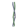
[English] 日本語
Yorodumi
- PDB-5ctd: Crystal structure of the type IX collagen NC2 hetero-trimerizatio... -

+
Open data


ID or keywords:

Loading...

-
Basic information

Entry
Database: PDB / ID: 5ctd
TitleCrystal structure of the type IX collagen NC2 hetero-trimerization domain with a guest fragment a2a1a1 of type I collagen
Components
  • Collagen alpha-1(I) chain,Collagen alpha-1(IX) chain
  • Collagen alpha-1(I) chain,Collagen alpha-3(IX) chain
  • Collagen alpha-2(I) chain,Collagen alpha-2(IX) chain
KeywordsSTRUCTURAL PROTEIN / collagen / hetero-trimerization / chain stagger / chain register / triple helix
Function / homology
Function and homology information


collagen type IX trimer / cellular response to fluoride / collagen type I trimer / tooth mineralization / protein heterotrimerization / cellular response to vitamin E / collagen type IV trimer / Anchoring fibril formation / Crosslinking of collagen fibrils / collagen biosynthetic process ...collagen type IX trimer / cellular response to fluoride / collagen type I trimer / tooth mineralization / protein heterotrimerization / cellular response to vitamin E / collagen type IV trimer / Anchoring fibril formation / Crosslinking of collagen fibrils / collagen biosynthetic process / Collagen chain trimerization / extracellular matrix assembly / Enhanced cleavage of VWF variant by ADAMTS13 / Defective VWF cleavage by ADAMTS13 variant / Defective VWF binding to collagen type I / platelet-derived growth factor binding / bone trabecula formation / extracellular matrix structural constituent conferring tensile strength / Enhanced binding of GP1BA variant to VWF multimer:collagen / Defective binding of VWF variant to GPIb:IX:V / intramembranous ossification / Extracellular matrix organization / embryonic skeletal system development / Collagen biosynthesis and modifying enzymes / cartilage development involved in endochondral bone morphogenesis / collagen metabolic process / skin morphogenesis / collagen-activated tyrosine kinase receptor signaling pathway / Signaling by PDGF / Platelet Adhesion to exposed collagen / endochondral ossification / NCAM1 interactions / cellular response to fibroblast growth factor stimulus / collagen fibril organization / negative regulation of cell-substrate adhesion / response to steroid hormone / face morphogenesis / odontogenesis / Scavenging by Class A Receptors / extracellular matrix structural constituent / skin development / MET activates PTK2 signaling / Assembly of collagen fibrils and other multimeric structures / Syndecan interactions / GP1b-IX-V activation signalling / blood vessel development / bone mineralization / SMAD binding / RUNX2 regulates osteoblast differentiation / Platelet Aggregation (Plug Formation) / Rho protein signal transduction / Collagen degradation / protein localization to nucleus / Non-integrin membrane-ECM interactions / ECM proteoglycans / response to hyperoxia / Integrin cell surface interactions / positive regulation of epithelial to mesenchymal transition / response to mechanical stimulus / cellular response to retinoic acid / GPVI-mediated activation cascade / cellular response to epidermal growth factor stimulus / response to cAMP / cellular response to transforming growth factor beta stimulus / visual perception / extracellular matrix organization / ossification / transforming growth factor beta receptor signaling pathway / secretory granule / skeletal system development / Cell surface interactions at the vascular wall / cellular response to glucose stimulus / sensory perception of sound / animal organ morphogenesis / cellular response to amino acid stimulus / SMAD2/SMAD3:SMAD4 heterotrimer regulates transcription / response to insulin / response to hydrogen peroxide / regulation of blood pressure / osteoblast differentiation / cellular response to mechanical stimulus / Immunoregulatory interactions between a Lymphoid and a non-Lymphoid cell / positive regulation of canonical Wnt signaling pathway / protein transport / response to estradiol / protein-macromolecule adaptor activity / cellular response to tumor necrosis factor / carbohydrate binding / Interleukin-4 and Interleukin-13 signaling / collagen-containing extracellular matrix / protease binding / positive regulation of cell migration / response to xenobiotic stimulus / endoplasmic reticulum lumen / positive regulation of DNA-templated transcription / protein homodimerization activity / extracellular space / extracellular exosome / extracellular region / identical protein binding
Similarity search - Function
: / Thrombospondin N-terminal -like domains. / Fibrillar collagen, C-terminal / Fibrillar collagen C-terminal domain / Fibrillar collagen C-terminal non-collagenous (NC1) domain profile. / Fibrillar collagens C-terminal domain / von Willebrand factor type C domain / VWFC domain signature. / VWFC domain profile. / von Willebrand factor (vWF) type C domain ...: / Thrombospondin N-terminal -like domains. / Fibrillar collagen, C-terminal / Fibrillar collagen C-terminal domain / Fibrillar collagen C-terminal non-collagenous (NC1) domain profile. / Fibrillar collagens C-terminal domain / von Willebrand factor type C domain / VWFC domain signature. / VWFC domain profile. / von Willebrand factor (vWF) type C domain / VWFC domain / Collagen triple helix repeat / Collagen triple helix repeat (20 copies) / Concanavalin A-like lectin/glucanase domain superfamily
Similarity search - Domain/homology
Collagen alpha-1(I) chain / Collagen alpha-2(I) chain / Collagen alpha-1(IX) chain / Collagen alpha-3(IX) chain / Collagen alpha-2(IX) chain
Similarity search - Component
Biological speciesHomo sapiens (human)
MethodX-RAY DIFFRACTION / SYNCHROTRON / MAD / Resolution: 1.5991 Å
AuthorsBoudko, S.P. / Bachinger, H.P.
Funding support United States, 1items
OrganizationGrant numberCountry
Shriners Hospitals for Children United States
CitationJournal: Sci Rep / Year: 2016
Title: Structural insight for chain selection and stagger control in collagen.
Authors: Boudko, S.P. / Bachinger, H.P.
History
DepositionJul 23, 2015Deposition site: RCSB / Processing site: RCSB
Revision 1.0Aug 3, 2016Provider: repository / Type: Initial release
Revision 1.1Jul 12, 2017Group: Database references / Derived calculations / Category: citation / pdbx_struct_oper_list
Item: _citation.country / _citation.journal_abbrev ..._citation.country / _citation.journal_abbrev / _citation.journal_id_CSD / _citation.journal_id_ISSN / _citation.journal_volume / _citation.page_first / _citation.page_last / _citation.pdbx_database_id_DOI / _citation.pdbx_database_id_PubMed / _citation.title / _citation.year / _pdbx_struct_oper_list.symmetry_operation

-
Structure visualization

Structure viewerMolecule:
MolmilJmol/JSmol

Downloads & links

-
Assembly

Deposited unit
A: Collagen alpha-1(I) chain,Collagen alpha-1(IX) chain
B: Collagen alpha-2(I) chain,Collagen alpha-2(IX) chain
C: Collagen alpha-1(I) chain,Collagen alpha-3(IX) chain


Theoretical massNumber of molelcules
Total (without water)21,0563
Polymers21,0563
Non-polymers00
Water3,495194
1


  • Idetical with deposited unit
  • defined by author&software
TypeNameSymmetry operationNumber
identity operation1_555x,y,z1
Buried area9230 Å2
ΔGint-69 kcal/mol
Surface area12800 Å2
MethodPISA
Unit cell
Length a, b, c (Å)27.890, 55.920, 62.100
Angle α, β, γ (deg.)90.00, 92.86, 90.00
Int Tables number4
Space group name H-MP1211
Symmetry operation#1: x,y,z
#2: -x,y+1/2,-z

-
Components

#1: Protein Collagen alpha-1(I) chain,Collagen alpha-1(IX) chain / Alpha-1 type I collagen


Mass: 7161.650 Da / Num. of mol.: 1 / Fragment: UNP Residues 572-583, UNP Residues 754-789
Source method: isolated from a genetically manipulated source
Source: (gene. exp.) Homo sapiens (human) / Gene: COL1A1, COL9A1 / Plasmid: pET22b(+) / Production host: Escherichia coli (E. coli) / Strain (production host): B834(DE3) / References: UniProt: P02452, UniProt: P20849
#2: Protein Collagen alpha-2(I) chain,Collagen alpha-2(IX) chain / Alpha-2 type I collagen


Mass: 6895.721 Da / Num. of mol.: 1 / Fragment: UNP Residues 484-495, UNP Residues 517-552
Source method: isolated from a genetically manipulated source
Source: (gene. exp.) Homo sapiens (human) / Gene: COL1A2, COL9A2 / Plasmid: pET22b(+) / Production host: Escherichia coli (E. coli) / Strain (production host): B834(DE3) / References: UniProt: P08123, UniProt: Q14055
#3: Protein Collagen alpha-1(I) chain,Collagen alpha-3(IX) chain / Alpha-1 type I collagen


Mass: 6999.000 Da / Num. of mol.: 1 / Fragment: UNP Residues 572-583, UNP Residues 517-553
Source method: isolated from a genetically manipulated source
Source: (gene. exp.) Homo sapiens (human) / Gene: COL1A1, COL9A3 / Plasmid: pET22b(+) / Production host: Escherichia coli (E. coli) / Strain (production host): B834(DE3) / References: UniProt: P02452, UniProt: Q14050
#4: Water ChemComp-HOH / water / Water


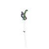
Mass: 18.015 Da / Num. of mol.: 194 / Source method: isolated from a natural source / Formula: H2O

-
Experimental details

-
Experiment

ExperimentMethod: X-RAY DIFFRACTION / Number of used crystals: 1

-
Sample preparation

CrystalDensity Matthews: 2.3 Å3/Da / Density % sol: 46.45 %
Crystal growTemperature: 295 K / Method: vapor diffusion, hanging drop / pH: 7.5 / Details: 0.1 M HEPES, 50 mM sodium acetate, 17% PEG 3,350

-
Data collection

DiffractionMean temperature: 100 K
Diffraction sourceSource: SYNCHROTRON / Site: ALS / Beamline: 4.2.2 / Wavelength: 0.97872, 0.97918, 0.96487
DetectorType: NOIR-1 / Detector: CCD / Date: Mar 23, 2014
RadiationMonochromator: Rosenbaum-Rock Si(111) sagitally focused monochromator
Protocol: MAD / Monochromatic (M) / Laue (L): M / Scattering type: x-ray
Radiation wavelength
IDWavelength (Å)Relative weight
10.978721
20.979181
30.964871
ReflectionRedundancy: 3.8 % / Number: 92373 / Rmerge(I) obs: 0.084 / Χ2: 1.56 / D res high: 1.6 Å / D res low: 50 Å / Num. obs: 24414 / % possible obs: 96.7
Diffraction reflection shell
Highest resolution (Å)Lowest resolution (Å)IDRmerge(I) obsChi squaredRedundancy
4.345010.0663.8153.7
3.454.3410.0671.6763.8
3.013.4510.0771.9423.9
2.743.0110.0691.873.9
2.542.7410.0821.9213.9
2.392.5410.0871.8533.9
2.272.3910.0831.6223.9
2.172.2710.0911.63.9
2.092.1710.0941.6423.9
2.022.0910.1041.5693.9
1.952.0210.1191.6433.9
1.91.9510.1361.3893.9
1.851.910.1681.2813.9
1.81.8510.2411.4673.9
1.761.810.2611.2033.9
1.721.7610.2750.9763.9
1.691.7210.2880.8833.8
1.661.6910.3210.7253.6
1.631.6610.3340.7873.3
1.61.6310.360.8233
ReflectionResolution: 1.599→50 Å / Num. obs: 24414 / % possible obs: 96.7 % / Redundancy: 3.8 % / Biso Wilson estimate: 16.2502146055 Å2 / Rmerge(I) obs: 0.084 / Rpim(I) all: 0.049 / Rrim(I) all: 0.097 / Χ2: 1.562 / Net I/av σ(I): 17.13 / Net I/σ(I): 15.6 / Num. measured all: 92373
Reflection shell

Diffraction-ID: 1 / Rejects: 0

Resolution (Å)Redundancy (%)Rmerge(I) obsNum. unique allCC1/2Rpim(I) allRrim(I) allΧ2% possible all
1.599-1.6330.3610950.770.2410.4350.82388.3
1.63-1.663.30.33411650.8970.2120.3970.78793.3
1.66-1.693.60.32112060.9430.1950.3760.72596.1
1.69-1.723.80.28812130.950.1710.3360.88395.7
1.72-1.763.90.27512090.9310.1620.320.97696
1.76-1.83.90.26112020.9460.1530.3031.20396.3
1.8-1.853.90.24112210.9550.1410.2791.46796.9
1.85-1.93.90.16812260.9760.0990.1951.28196.6
1.9-1.953.90.13612010.9760.080.1581.38997.2
1.95-2.023.90.11912400.9840.070.1381.64397.2
2.02-2.093.90.10412180.9880.0610.1211.56996.7
2.09-2.173.90.09412330.990.0550.1091.64297.9
2.17-2.273.90.09112220.9910.0530.1051.697.7
2.27-2.393.90.08312390.9920.0490.0971.62297.6
2.39-2.543.90.08712530.990.0510.1011.85398.5
2.54-2.743.90.08212310.9940.0480.0951.92198.4
2.74-3.013.90.06912410.9960.0410.0811.8798.2
3.01-3.453.90.07712680.9910.0450.0891.94298.9
3.45-4.343.80.06712550.9930.040.0781.67698.9
4.34-503.70.06612760.9940.0390.0773.81596.8

-
Phasing

PhasingMethod: MAD
Phasing set
ID
1
2
3
Phasing MADD res high: 1.6 Å / D res low: 1000 Å / FOM : 0.4 / Reflection: 24386
Phasing MAD set
Clust-IDExpt-IDSet-IDWavelength (Å)F double prime refinedF prime refined
3 wavelength110.97875.02-6.87
3 wavelength120.97923.2-8.06
3 wavelength130.96493.76-5.17
Phasing MAD set site
IDCartn x (Å)Cartn y (Å)Cartn z (Å)Atom type symbolB isoOccupancy
19.71328.66527.792SE19.11.86
24.33955.53727.465SE15.21.33
314.9247.16329.568SE25.71.33
Phasing MAD shell
Resolution (Å)FOM Reflection
5.84-10000.71125
3.66-5.840.532050
2.85-3.660.582615
2.41-2.850.573088
2.13-2.410.53445
1.93-2.130.373806
1.77-1.930.234112
1.65-1.770.144145

-
Processing

Software
NameVersionClassification
PHENIX1.9_1692refinement
HKL-2000data collection
HKL-2000data scaling
SOLVEphasing
PDB_EXTRACT3.15data extraction
HKL-2000data reduction
RefinementMethod to determine structure: MAD / Resolution: 1.5991→41.532 Å / SU ML: 0.17 / Cross valid method: THROUGHOUT / σ(F): 0 / Phase error: 21.2 / Stereochemistry target values: MLHL
RfactorNum. reflection% reflection
Rfree0.2025 1962 8.22 %
Rwork0.1771 --
obs0.1791 23880 94.36 %
Solvent computationShrinkage radii: 0.9 Å / VDW probe radii: 1.11 Å / Solvent model: FLAT BULK SOLVENT MODEL
Refinement stepCycle: LAST / Resolution: 1.5991→41.532 Å
ProteinNucleic acidLigandSolventTotal
Num. atoms1416 0 0 194 1610
Refine LS restraints
Refine-IDTypeDev idealNumber
X-RAY DIFFRACTIONf_bond_d0.0121497
X-RAY DIFFRACTIONf_angle_d1.3832056
X-RAY DIFFRACTIONf_dihedral_angle_d11.967603
X-RAY DIFFRACTIONf_chiral_restr0.046197
X-RAY DIFFRACTIONf_plane_restr0.007305
LS refinement shell
Resolution (Å)Rfactor RfreeNum. reflection RfreeRfactor RworkNum. reflection RworkRefine-ID% reflection obs (%)
1.5991-1.63910.27071140.27081319X-RAY DIFFRACTION78
1.6391-1.68340.26841390.2451429X-RAY DIFFRACTION89
1.6834-1.73290.25881400.22231514X-RAY DIFFRACTION91
1.7329-1.78880.25481250.22741532X-RAY DIFFRACTION94
1.7888-1.85280.27631470.21011553X-RAY DIFFRACTION94
1.8528-1.9270.23451380.19751558X-RAY DIFFRACTION95
1.927-2.01470.21471440.18161606X-RAY DIFFRACTION96
2.0147-2.12090.18991490.17551579X-RAY DIFFRACTION97
2.1209-2.25370.18571360.16271603X-RAY DIFFRACTION97
2.2537-2.42770.17571520.16391620X-RAY DIFFRACTION98
2.4277-2.6720.18081420.16981645X-RAY DIFFRACTION98
2.672-3.05860.18841380.16561638X-RAY DIFFRACTION98
3.0586-3.8530.19081490.16951643X-RAY DIFFRACTION99
3.853-41.54580.19581490.16171679X-RAY DIFFRACTION98

+
About Yorodumi

-
News

-
Feb 9, 2022. New format data for meta-information of EMDB entries

New format data for meta-information of EMDB entries

  • Version 3 of the EMDB header file is now the official format.
  • The previous official version 1.9 will be removed from the archive.

Related info.:EMDB header

External links:wwPDB to switch to version 3 of the EMDB data model

-
Aug 12, 2020. Covid-19 info

Covid-19 info

URL: https://pdbj.org/emnavi/covid19.php

New page: Covid-19 featured information page in EM Navigator.

Related info.:Covid-19 info / Mar 5, 2020. Novel coronavirus structure data

+
Mar 5, 2020. Novel coronavirus structure data

Novel coronavirus structure data

Related info.:Yorodumi Speices / Aug 12, 2020. Covid-19 info

External links:COVID-19 featured content - PDBj / Molecule of the Month (242):Coronavirus Proteases

+
Jan 31, 2019. EMDB accession codes are about to change! (news from PDBe EMDB page)

EMDB accession codes are about to change! (news from PDBe EMDB page)

  • The allocation of 4 digits for EMDB accession codes will soon come to an end. Whilst these codes will remain in use, new EMDB accession codes will include an additional digit and will expand incrementally as the available range of codes is exhausted. The current 4-digit format prefixed with “EMD-” (i.e. EMD-XXXX) will advance to a 5-digit format (i.e. EMD-XXXXX), and so on. It is currently estimated that the 4-digit codes will be depleted around Spring 2019, at which point the 5-digit format will come into force.
  • The EM Navigator/Yorodumi systems omit the EMD- prefix.

Related info.:Q: What is EMD? / ID/Accession-code notation in Yorodumi/EM Navigator

External links:EMDB Accession Codes are Changing Soon! / Contact to PDBj

+
Jul 12, 2017. Major update of PDB

Major update of PDB

  • wwPDB released updated PDB data conforming to the new PDBx/mmCIF dictionary.
  • This is a major update changing the version number from 4 to 5, and with Remediation, in which all the entries are updated.
  • In this update, many items about electron microscopy experimental information are reorganized (e.g. em_software).
  • Now, EM Navigator and Yorodumi are based on the updated data.

External links:wwPDB Remediation / Enriched Model Files Conforming to OneDep Data Standards Now Available in the PDB FTP Archive

-
Yorodumi

Thousand views of thousand structures

  • Yorodumi is a browser for structure data from EMDB, PDB, SASBDB, etc.
  • This page is also the successor to EM Navigator detail page, and also detail information page/front-end page for Omokage search.
  • The word "yorodu" (or yorozu) is an old Japanese word meaning "ten thousand". "mi" (miru) is to see.

Related info.:EMDB / PDB / SASBDB / Comparison of 3 databanks / Yorodumi Search / Aug 31, 2016. New EM Navigator & Yorodumi / Yorodumi Papers / Jmol/JSmol / Function and homology information / Changes in new EM Navigator and Yorodumi

Read more