 Movie
Movie Controller


+ Open data

- Basic information
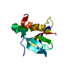
| Entry | Database: PDB / ID: 5chy | ||||||
|---|---|---|---|---|---|---|---|
| Title | STRUCTURE OF CHEMOTAXIS PROTEIN CHEY | ||||||
 Components Components | CHEY | ||||||
 Keywords Keywords | SIGNAL TRANSDUCTION PROTEIN / RESPONSE REGULATORS / TWO-COMPONENT SYSTEMS | ||||||
| Function / homology |  Function and homology information Function and homology informationbacterial-type flagellum basal body, C ring / bacterial-type flagellum rotor complex / bacterial-type flagellum-dependent swimming motility / aerotaxis / regulation of bacterial-type flagellum-dependent cell motility / regulation of chemotaxis / internal peptidyl-lysine acetylation / thermotaxis / bacterial-type flagellum / phosphorelay response regulator activity ...bacterial-type flagellum basal body, C ring / bacterial-type flagellum rotor complex / bacterial-type flagellum-dependent swimming motility / aerotaxis / regulation of bacterial-type flagellum-dependent cell motility / regulation of chemotaxis / internal peptidyl-lysine acetylation / thermotaxis / bacterial-type flagellum / phosphorelay response regulator activity / protein acetylation / acetyltransferase activity / phosphorelay signal transduction system / chemotaxis / magnesium ion binding / signal transduction / cytoplasm / cytosol Similarity search - Function | ||||||
| Biological species |  | ||||||
| Method |  X-RAY DIFFRACTION / MOLECULAR REPLACEMENT / Resolution: 2 Å X-RAY DIFFRACTION / MOLECULAR REPLACEMENT / Resolution: 2 Å | ||||||
 Authors Authors | Zhu, X. / Rebello, J. / Matsumura, P. / Volz, K. | ||||||
 Citation Citation | Journal: J.Biol.Chem. / Year: 1997 Title: Crystal structures of CheY mutants Y106W and T87I/Y106W. CheY activation correlates with movement of residue 106. Authors: Zhu, X. / Rebello, J. / Matsumura, P. / Volz, K. #1: Journal: J.Bacteriol. / Year: 1996Title: Tyrosine 106 Plays an Important Role in Chemotaxis Signal Transduction in Escherichia Coli Authors: Zhu, X. / Amsler, C.D. / Volz, K. / Matsumura, P. #2: Journal: J.Biol.Chem. / Year: 1995Title: Uncoupled Phosphorylation and Activation in Bacterial Chemotaxis. The 2.1-A Structure of a Threonine to Isoleucine Mutant at Position 87 of Chey Authors: Ganguli, S. / Wang, H. / Matsumura, P. / Volz, K. #3: Journal: Biochemistry / Year: 1993Title: Structural Conservation in the Chey Superfamily Authors: Volz, K. #4: Journal: J.Biol.Chem. / Year: 1991Title: Crystal Structure of Escherichia Coli Chey Refined at 1.7-A Resolution Authors: Volz, K. / Matsumura, P. | ||||||
| History |
|
- Structure visualization
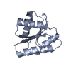
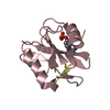
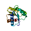
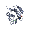
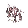
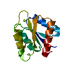
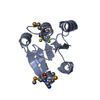
| Structure viewer | Molecule: MolmilJmol/JSmol |
|---|
- Downloads & links
-Download
| PDBx/mmCIF format | 5chy.cif.gz | 38.3 KB | Display | PDBx/mmCIF format |
|---|---|---|---|---|
| PDB format | pdb5chy.ent.gz | 26.2 KB | Display | PDB format |
| PDBx/mmJSON format | 5chy.json.gz | Tree view | PDBx/mmJSON format | |
| Others |  Other downloads Other downloads |
-Validation report
| Arichive directory | https://data.pdbj.org/pub/pdb/validation_reports/ch/5chyftp://data.pdbj.org/pub/pdb/validation_reports/ch/5chy | HTTPS FTP |
|---|
-Related structure data


-Links
 PDBj
PDBj

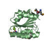
- Assembly
| Deposited unit | 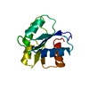
| ||||||||
|---|---|---|---|---|---|---|---|---|---|
| 1 |
| ||||||||
| Unit cell |
|
-Components
| #1: Protein | Mass: 14004.171 Da / Num. of mol.: 1 / Mutation: Y106W Source method: isolated from a genetically manipulated source Source: (gene. exp.) |
|---|---|
| #2: Chemical | ChemComp-CA /   Mass: 40.078 Da / Num. of mol.: 1 / Source method: obtained synthetically / Formula: Ca Mass: 40.078 Da / Num. of mol.: 1 / Source method: obtained synthetically / Formula: Ca |
| #3: Water | ChemComp-HOH /  Mass: 18.015 Da / Num. of mol.: 83 / Source method: isolated from a natural source / Formula: H2O Mass: 18.015 Da / Num. of mol.: 83 / Source method: isolated from a natural source / Formula: H2O |
-Experimental details
-Experiment
| Experiment | Method: X-RAY DIFFRACTION / Number of used crystals: 1 |
|---|
- Sample preparation
| Crystal | Density Matthews: 2.02 Å3/Da / Density % sol: 39.12 % | ||||||||||||||||||||
|---|---|---|---|---|---|---|---|---|---|---|---|---|---|---|---|---|---|---|---|---|---|
| Crystal grow | pH: 6 Details: 28% PEG 8000, 0.12 M CALCIUM ACETATE, 0.1 M CACODYLATE BUFFER, PH 6.0 | ||||||||||||||||||||
| Crystal grow | *PLUS Temperature: 4 ℃ / Method: vapor diffusion, hanging drop | ||||||||||||||||||||
| Components of the solutions | *PLUS
|
-Data collection
| Diffraction | Mean temperature: 292 K |
|---|---|
| Diffraction source | Source: ROTATING ANODE / Wavelength: 1.5418 |
| Detector | Type: RIGAKU RAXIS II / Detector: IMAGE PLATE |
| Radiation | Monochromatic (M) / Laue (L): M / Scattering type: x-ray |
| Radiation wavelength | Wavelength: 1.5418 Å / Relative weight: 1 |
| Reflection | Highest resolution: 2 Å / Num. obs: 7433 / % possible obs: 92 % / Observed criterion σ(I): 1 / Rmerge(I) obs: 0.066 / Net I/σ(I): 1 |
| Reflection shell | Resolution: 2→2.13 Å / % possible all: 80.3 |
| Reflection | *PLUS Num. measured all: 21022 |
| Reflection shell | *PLUS % possible obs: 80.3 % |
- Processing
| Software |
| ||||||||||||||||||||||||||||||||||||||||||||||||||||||||||||||||||||||||||||||||||||
|---|---|---|---|---|---|---|---|---|---|---|---|---|---|---|---|---|---|---|---|---|---|---|---|---|---|---|---|---|---|---|---|---|---|---|---|---|---|---|---|---|---|---|---|---|---|---|---|---|---|---|---|---|---|---|---|---|---|---|---|---|---|---|---|---|---|---|---|---|---|---|---|---|---|---|---|---|---|---|---|---|---|---|---|---|---|
| Refinement | Method to determine structure: MOLECULAR REPLACEMENT / Resolution: 2→10 Å / σ(F): 2
| ||||||||||||||||||||||||||||||||||||||||||||||||||||||||||||||||||||||||||||||||||||
| Displacement parameters | Biso mean: 24.5 Å2 | ||||||||||||||||||||||||||||||||||||||||||||||||||||||||||||||||||||||||||||||||||||
| Refinement step | Cycle: LAST / Resolution: 2→10 Å
| ||||||||||||||||||||||||||||||||||||||||||||||||||||||||||||||||||||||||||||||||||||
| Refine LS restraints |
| ||||||||||||||||||||||||||||||||||||||||||||||||||||||||||||||||||||||||||||||||||||
| Software | *PLUS Name: PROFFT / Classification: refinement | ||||||||||||||||||||||||||||||||||||||||||||||||||||||||||||||||||||||||||||||||||||
| Refinement | *PLUS Rfactor obs: 0.185 | ||||||||||||||||||||||||||||||||||||||||||||||||||||||||||||||||||||||||||||||||||||
| Solvent computation | *PLUS | ||||||||||||||||||||||||||||||||||||||||||||||||||||||||||||||||||||||||||||||||||||
| Displacement parameters | *PLUS | ||||||||||||||||||||||||||||||||||||||||||||||||||||||||||||||||||||||||||||||||||||
| LS refinement shell | *PLUS Highest resolution: 2 Å / Lowest resolution: 2.13 Å / Total num. of bins used: 7 / Num. reflection obs: 1357 / Rfactor obs: 0.278 |
