 Movie
Movie Controller

[English] 日本語
 Yorodumi
- PDB-5che: Crystal structure of Arabidopsis glutamyl-tRNA reductase in compl... -
+ Open data

- Basic information
| Entry | Database: PDB / ID: 5che | ||||||
|---|---|---|---|---|---|---|---|
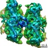
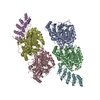
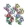
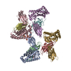
| Title | Crystal structure of Arabidopsis glutamyl-tRNA reductase in complex with its regulatory proteins | ||||||
 Components Components |
| ||||||
 Keywords Keywords | OXIDOREDUCTASE / GluTR / tertiary complex / regulatory proteins / anchor protein | ||||||
| Function / homology |  Function and homology information Function and homology informationglutamyl-tRNA reductase / glutamyl-tRNA reductase activity / tetrapyrrole biosynthetic process / positive regulation of heme biosynthetic process / chlorophyll biosynthetic process / porphyrin-containing compound biosynthetic process / chloroplast membrane / oxylipin biosynthetic process / chloroplast thylakoid / : ...glutamyl-tRNA reductase / glutamyl-tRNA reductase activity / tetrapyrrole biosynthetic process / positive regulation of heme biosynthetic process / chlorophyll biosynthetic process / porphyrin-containing compound biosynthetic process / chloroplast membrane / oxylipin biosynthetic process / chloroplast thylakoid / : / chloroplast envelope / photosynthetic electron transport chain / heme biosynthetic process / chloroplast stroma / chloroplast thylakoid membrane / response to light stimulus / protein-membrane adaptor activity / chloroplast / post-embryonic development / NADP binding / protein-containing complex / cytosol Similarity search - Function | ||||||
| Biological species |  | ||||||
| Method |  X-RAY DIFFRACTION / SYNCHROTRON / MOLECULAR REPLACEMENT / molecular replacement / Resolution: 3.203 Å X-RAY DIFFRACTION / SYNCHROTRON / MOLECULAR REPLACEMENT / molecular replacement / Resolution: 3.203 Å | ||||||
 Authors Authors | Fang, Y. / Liu, L. | ||||||
 Citation Citation | Journal: Sci Rep / Year: 2016 Title: The Arabidopsis glutamyl-tRNA reductase (GluTR) forms a ternary complex with FLU and GluTR-binding protein Authors: Fang, Y. / Zhao, S. / Zhang, F. / Zhao, A. / Zhang, W. / Zhang, M. / Liu, L. | ||||||
| History |
|
- Structure visualization
| Structure viewer | Molecule: MolmilJmol/JSmol |
|---|
- Downloads & links
-Download
| PDBx/mmCIF format | 5che.cif.gz | 317.6 KB | Display | PDBx/mmCIF format |
|---|---|---|---|---|
| PDB format | pdb5che.ent.gz | 252.6 KB | Display | PDB format |
| PDBx/mmJSON format | 5che.json.gz | Tree view | PDBx/mmJSON format | |
| Others |  Other downloads Other downloads |
-Validation report
| Arichive directory | https://data.pdbj.org/pub/pdb/validation_reports/ch/5cheftp://data.pdbj.org/pub/pdb/validation_reports/ch/5che | HTTPS FTP |
|---|
-Related structure data


-Links
 PDBj
PDBj


- Assembly
| Deposited unit | 
| ||||||||
|---|---|---|---|---|---|---|---|---|---|
| 1 |
| ||||||||
| Unit cell |
|
-Components
| #1: Protein | Mass: 52139.902 Da / Num. of mol.: 2 / Fragment: UNP residues 73-543 Source method: isolated from a genetically manipulated source Source: (gene. exp.)  #2: Protein | Mass: 34272.648 Da / Num. of mol.: 2 / Fragment: UNP residues 42-317 Source method: isolated from a genetically manipulated source Source: (gene. exp.) #3: Protein | Mass: 17771.264 Da / Num. of mol.: 2 / Fragment: UNP residues 195-316 Source method: isolated from a genetically manipulated source Source: (gene. exp.) #4: Water | ChemComp-HOH / |  Mass: 18.015 Da / Num. of mol.: 6 / Source method: isolated from a natural source / Formula: H2O Mass: 18.015 Da / Num. of mol.: 6 / Source method: isolated from a natural source / Formula: H2O |
|---|
-Experimental details
-Experiment
| Experiment | Method: X-RAY DIFFRACTION / Number of used crystals: 1 |
|---|
- Sample preparation
| Crystal | Density Matthews: 2.68 Å3/Da / Density % sol: 54.09 % |
|---|---|
| Crystal grow | Temperature: 289 K / Method: evaporation / pH: 7 / Details: PEG 3350, sodium malonate, LiCl, MPD |
-Data collection
| Diffraction | Mean temperature: 100 K |
|---|---|
| Diffraction source | Source: SYNCHROTRON / Site: SSRF  / Beamline: BL17U / Wavelength: 0.9793 Å / Beamline: BL17U / Wavelength: 0.9793 Å |
| Detector | Type: RAYONIX MX-325 / Detector: CCD / Date: Jan 19, 2015 |
| Radiation | Protocol: SINGLE WAVELENGTH / Monochromatic (M) / Laue (L): M / Scattering type: x-ray |
| Radiation wavelength | Wavelength: 0.9793 Å / Relative weight: 1 |
| Reflection | Resolution: 3.2→50 Å / Num. all: 36775 / Num. obs: 36775 / % possible obs: 99.6 % / Redundancy: 4.3 % / Rmerge(I) obs: 0.132 / Net I/σ(I): 10 |
| Reflection shell | Resolution: 3.2→3.31 Å / Redundancy: 4.3 % / Rmerge(I) obs: 0.831 / Mean I/σ(I) obs: 1.68 / % possible all: 99.9 |
-Phasing
| Phasing | Method: molecular replacement |
|---|
- Processing
| Software |
| |||||||||||||||||||||||||||||||||||||||||||||||||||||||||||||||||||||||||||||||||||||||||||
|---|---|---|---|---|---|---|---|---|---|---|---|---|---|---|---|---|---|---|---|---|---|---|---|---|---|---|---|---|---|---|---|---|---|---|---|---|---|---|---|---|---|---|---|---|---|---|---|---|---|---|---|---|---|---|---|---|---|---|---|---|---|---|---|---|---|---|---|---|---|---|---|---|---|---|---|---|---|---|---|---|---|---|---|---|---|---|---|---|---|---|---|---|
| Refinement | Method to determine structure: MOLECULAR REPLACEMENT Starting model: 4N7R, 4YVQ Resolution: 3.203→39.265 Å / FOM work R set: 0.7788 / SU ML: 0.45 / Cross valid method: FREE R-VALUE / σ(F): 1.34 / Phase error: 28.67 / Stereochemistry target values: ML
| |||||||||||||||||||||||||||||||||||||||||||||||||||||||||||||||||||||||||||||||||||||||||||
| Solvent computation | Shrinkage radii: 0.9 Å / VDW probe radii: 1.11 Å / Solvent model: FLAT BULK SOLVENT MODEL | |||||||||||||||||||||||||||||||||||||||||||||||||||||||||||||||||||||||||||||||||||||||||||
| Displacement parameters | Biso max: 108.18 Å2 / Biso mean: 26.75 Å2 / Biso min: 1.43 Å2 | |||||||||||||||||||||||||||||||||||||||||||||||||||||||||||||||||||||||||||||||||||||||||||
| Refinement step | Cycle: final / Resolution: 3.203→39.265 Å
| |||||||||||||||||||||||||||||||||||||||||||||||||||||||||||||||||||||||||||||||||||||||||||
| Refine LS restraints |
| |||||||||||||||||||||||||||||||||||||||||||||||||||||||||||||||||||||||||||||||||||||||||||
| LS refinement shell | Refine-ID: X-RAY DIFFRACTION / Total num. of bins used: 12
|
