 Movie
Movie Controller

[English] 日本語
 Yorodumi
- PDB-4yvq: Crystal Structure of FLU-TPR in Complex with the C-terminal Regio... -
+ Open data

- Basic information
| Entry | Database: PDB / ID: 4yvq | ||||||
|---|---|---|---|---|---|---|---|
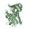
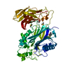
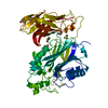
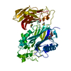
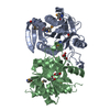
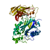
| Title | Crystal Structure of FLU-TPR in Complex with the C-terminal Region of GluTR | ||||||
 Components Components |
| ||||||
 Keywords Keywords | OXIDOREDUCTASE/FLUORESCENT PROTEIN / TPR / chloroplast / protein-protein interaction / OXIDOREDUCTASE-FLUORESCENT PROTEIN complex | ||||||
| Function / homology |  Function and homology information Function and homology informationglutamyl-tRNA reductase / glutamyl-tRNA reductase activity / chlorophyll biosynthetic process / porphyrin-containing compound biosynthetic process / chloroplast membrane / oxylipin biosynthetic process / chloroplast thylakoid / : / chloroplast envelope / heme biosynthetic process ...glutamyl-tRNA reductase / glutamyl-tRNA reductase activity / chlorophyll biosynthetic process / porphyrin-containing compound biosynthetic process / chloroplast membrane / oxylipin biosynthetic process / chloroplast thylakoid / : / chloroplast envelope / heme biosynthetic process / chloroplast thylakoid membrane / response to light stimulus / chloroplast / NADP binding / protein-containing complex / cytosol Similarity search - Function | ||||||
| Biological species |  | ||||||
| Method |  X-RAY DIFFRACTION / SYNCHROTRON / MOLECULAR REPLACEMENT / Resolution: 2.401 Å X-RAY DIFFRACTION / SYNCHROTRON / MOLECULAR REPLACEMENT / Resolution: 2.401 Å | ||||||
 Authors Authors | Zhang, M. / Zhang, F. / Liu, L. | ||||||
 Citation Citation | Journal: J.Biol.Chem. / Year: 2015 Title: The Non-canonical Tetratricopeptide Repeat (TPR) Domain of Fluorescent (FLU) Mediates Complex Formation with Glutamyl-tRNA Reductase. Authors: Zhang, M. / Zhang, F. / Fang, Y. / Chen, X. / Chen, Y. / Zhang, W. / Dai, H.E. / Lin, R. / Liu, L. | ||||||
| History |
|
- Structure visualization
| Structure viewer | Molecule: MolmilJmol/JSmol |
|---|
- Downloads & links
-Download
| PDBx/mmCIF format | 4yvq.cif.gz | 100.5 KB | Display | PDBx/mmCIF format |
|---|---|---|---|---|
| PDB format | pdb4yvq.ent.gz | 75.8 KB | Display | PDB format |
| PDBx/mmJSON format | 4yvq.json.gz | Tree view | PDBx/mmJSON format | |
| Others |  Other downloads Other downloads |
-Validation report
| Arichive directory | https://data.pdbj.org/pub/pdb/validation_reports/yv/4yvqftp://data.pdbj.org/pub/pdb/validation_reports/yv/4yvq | HTTPS FTP |
|---|
-Related structure data
| Related structure data |  4yvoSC S: Starting model for refinement C: citing same article ( |
|---|---|
| Similar structure data |


-Links
 PDBj
PDBj


- Assembly
| Deposited unit | 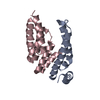
| ||||||||
|---|---|---|---|---|---|---|---|---|---|
| 1 | 
| ||||||||
| Unit cell |
|
-Components
| #1: Protein | Mass: 12142.102 Da / Num. of mol.: 1 / Fragment: C-terminal Region (UNP RESIDUES 440-543) Source method: isolated from a genetically manipulated source Source: (gene. exp.)  |
|---|---|
| #2: Protein | Mass: 17771.264 Da / Num. of mol.: 1 / Fragment: TPR Domain (UNP RESIDUES 195-316) Source method: isolated from a genetically manipulated source Source: (gene. exp.) |
| #3: Water | ChemComp-HOH /  Mass: 18.015 Da / Num. of mol.: 81 / Source method: isolated from a natural source / Formula: H2O Mass: 18.015 Da / Num. of mol.: 81 / Source method: isolated from a natural source / Formula: H2O |
-Experimental details
-Experiment
| Experiment | Method: X-RAY DIFFRACTION / Number of used crystals: 1 |
|---|
- Sample preparation
| Crystal | Density Matthews: 2.18 Å3/Da / Density % sol: 43.54 % |
|---|---|
| Crystal grow | Temperature: 277 K / Method: evaporation Details: 0.15M KBr, 30% w/v Polyethylene glycol monomethyl ether 2000 |
-Data collection
| Diffraction | Mean temperature: 100 K |
|---|---|
| Diffraction source | Source: SYNCHROTRON / Site: SSRF  / Beamline: BL17U / Wavelength: 0.9793 Å / Beamline: BL17U / Wavelength: 0.9793 Å |
| Detector | Type: ADSC QUANTUM 315r / Detector: CCD / Date: Nov 30, 2013 |
| Radiation | Monochromator: double crystal / Protocol: SINGLE WAVELENGTH / Monochromatic (M) / Laue (L): M / Scattering type: x-ray |
| Radiation wavelength | Wavelength: 0.9793 Å / Relative weight: 1 |
| Reflection | Resolution: 2.4→50 Å / Num. obs: 11100 / % possible obs: 99.9 % / Redundancy: 10.2 % / Net I/σ(I): 26.6 |
- Processing
| Software |
| ||||||||||||||||||||||||||||||||||||||||
|---|---|---|---|---|---|---|---|---|---|---|---|---|---|---|---|---|---|---|---|---|---|---|---|---|---|---|---|---|---|---|---|---|---|---|---|---|---|---|---|---|---|
| Refinement | Method to determine structure: MOLECULAR REPLACEMENT Starting model: 4YVO Resolution: 2.401→34.28 Å / FOM work R set: 0.8144 / SU ML: 0.25 / Cross valid method: FREE R-VALUE / σ(F): 1.35 / Phase error: 23.74 / Stereochemistry target values: ML
| ||||||||||||||||||||||||||||||||||||||||
| Solvent computation | Shrinkage radii: 0.9 Å / VDW probe radii: 1.11 Å / Solvent model: FLAT BULK SOLVENT MODEL | ||||||||||||||||||||||||||||||||||||||||
| Displacement parameters | Biso max: 130.65 Å2 / Biso mean: 48.66 Å2 / Biso min: 22.42 Å2 | ||||||||||||||||||||||||||||||||||||||||
| Refinement step | Cycle: final / Resolution: 2.401→34.28 Å
| ||||||||||||||||||||||||||||||||||||||||
| Refine LS restraints |
| ||||||||||||||||||||||||||||||||||||||||
| LS refinement shell | Refine-ID: X-RAY DIFFRACTION / Total num. of bins used: 4 / % reflection obs: 100 %
| ||||||||||||||||||||||||||||||||||||||||
| Refinement TLS params. | Method: refined / Origin x: 32.767 Å / Origin y: 39.4155 Å / Origin z: 5.3824 Å
| ||||||||||||||||||||||||||||||||||||||||
| Refinement TLS group | Selection details: all |
