 Movie
Movie Controller


+ Open data

- Basic information
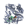
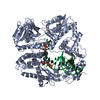
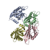
| Entry | Database: PDB / ID: 5ch6 | ||||||
|---|---|---|---|---|---|---|---|
| Title | Crystal Structure of FRIGIDA Flowering-time Regulator | ||||||
 Components Components | FRIGIDA | ||||||
 Keywords Keywords | TRANSCRIPTION / Flowering / Epigenetics / Transcription activator | ||||||
| Function / homology | Frigida-like / Frigida-like protein / flower development / cell differentiation / FRIGIDA-like protein Function and homology information Function and homology information | ||||||
| Biological species |  | ||||||
| Method |  X-RAY DIFFRACTION / SYNCHROTRON / SAD / Resolution: 3.26 Å X-RAY DIFFRACTION / SYNCHROTRON / SAD / Resolution: 3.26 Å | ||||||
 Authors Authors | Hyun, K. / Song, J.J. | ||||||
| Funding support |  Korea, Republic Of, 1items Korea, Republic Of, 1items
| ||||||
 Citation Citation | Journal: Mol Plant / Year: 2016 Title: Structural Analysis of FRIGIDA Flowering-Time Regulator Authors: Hyun, K. / Oh, J.E. / Park, J. / Noh, Y.S. / Song, J.J. | ||||||
| History |
|
- Structure visualization
| Structure viewer | Molecule: MolmilJmol/JSmol |
|---|
- Downloads & links
-Download
| PDBx/mmCIF format | 5ch6.cif.gz | 175.6 KB | Display | PDBx/mmCIF format |
|---|---|---|---|---|
| PDB format | pdb5ch6.ent.gz | 142.3 KB | Display | PDB format |
| PDBx/mmJSON format | 5ch6.json.gz | Tree view | PDBx/mmJSON format | |
| Others |  Other downloads Other downloads |
-Validation report
| Arichive directory | https://data.pdbj.org/pub/pdb/validation_reports/ch/5ch6ftp://data.pdbj.org/pub/pdb/validation_reports/ch/5ch6 | HTTPS FTP |
|---|
-Related structure data
| Similar structure data |
|---|


-Links
 PDBj
PDBj- Assembly
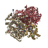
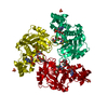
| Deposited unit | 
| ||||||||
|---|---|---|---|---|---|---|---|---|---|
| 1 |
| ||||||||
| Unit cell |
|
-Components
| #1: Protein | Mass: 32854.285 Da / Num. of mol.: 3 Source method: isolated from a genetically manipulated source Source: (gene. exp.)  Sequence details | SEQUENCE OF THE PROTEIN IS BASED ON DATABASE XP_002283789.1, AND NOT AVAILABLE FROM UNIPROT AT THE ...SEQUENCE OF THE PROTEIN IS BASED ON DATABASE XP_002283789.1, AND NOT AVAILABLE FROM UNIPROT AT THE TIME OF PROCESSING | |
|---|
-Experimental details
-Experiment
| Experiment | Method: X-RAY DIFFRACTION / Number of used crystals: 1 |
|---|
- Sample preparation
| Crystal | Density Matthews: 7.37447 Å3/Da / Density % sol: 83.320839 % |
|---|---|
| Crystal grow | Temperature: 293 K / Method: vapor diffusion, hanging drop / pH: 4.6 / Details: sodium formate, sodium acetate trihydrate pH 4.6 |
-Data collection
| Diffraction | Mean temperature: 293 K |
|---|---|
| Diffraction source | Source: SYNCHROTRON / Site: Photon Factory  / Beamline: BL-5A / Wavelength: 1 Å / Beamline: BL-5A / Wavelength: 1 Å |
| Detector | Type: ADSC QUANTUM 315 / Detector: CCD / Date: Mar 26, 2012 |
| Radiation | Protocol: SINGLE WAVELENGTH / Monochromatic (M) / Laue (L): M / Scattering type: x-ray |
| Radiation wavelength | Wavelength: 1 Å / Relative weight: 1 |
| Reflection | Resolution: 3.25→50 Å / Num. obs: 43466 / % possible obs: 92.6 % / Redundancy: 4.7 % / Net I/σ(I): 27.24 |
- Processing
| Software |
| ||||||||||||||||||||||||||||||||||||||||||||||||||||||||||||
|---|---|---|---|---|---|---|---|---|---|---|---|---|---|---|---|---|---|---|---|---|---|---|---|---|---|---|---|---|---|---|---|---|---|---|---|---|---|---|---|---|---|---|---|---|---|---|---|---|---|---|---|---|---|---|---|---|---|---|---|---|---|
| Refinement | Method to determine structure: SAD / Resolution: 3.26→46.96 Å / Rfactor Rfree error: 0.004 / Data cutoff high absF: 160159.34 / Data cutoff low absF: 0 / Isotropic thermal model: RESTRAINED / Cross valid method: THROUGHOUT / σ(F): 0 / Details: BULK SOLVENT MODEL USED
| ||||||||||||||||||||||||||||||||||||||||||||||||||||||||||||
| Solvent computation | Solvent model: FLAT MODEL / Bsol: 85.1672 Å2 / ksol: 0.32 e/Å3 | ||||||||||||||||||||||||||||||||||||||||||||||||||||||||||||
| Displacement parameters | Biso mean: 121.4 Å2
| ||||||||||||||||||||||||||||||||||||||||||||||||||||||||||||
| Refine analyze |
| ||||||||||||||||||||||||||||||||||||||||||||||||||||||||||||
| Refinement step | Cycle: 1 / Resolution: 3.26→46.96 Å /
| ||||||||||||||||||||||||||||||||||||||||||||||||||||||||||||
| Refine LS restraints |
| ||||||||||||||||||||||||||||||||||||||||||||||||||||||||||||
| LS refinement shell | Resolution: 3.25→3.45 Å / Rfactor Rfree error: 0.107 / Total num. of bins used: 6
| ||||||||||||||||||||||||||||||||||||||||||||||||||||||||||||
| Xplor file |
|
