Mass: 18.015 Da / Num. of mol.: 106 / Source method: isolated from a natural source / Formula: D2O
Nonpolymer details
AUTHOR STATES THAT THE PYRIDINE NITROGEN FOR THE LIGAND RIS IS IN THE PROTONATED FORM IN THE ...AUTHOR STATES THAT THE PYRIDINE NITROGEN FOR THE LIGAND RIS IS IN THE PROTONATED FORM IN THE CRYSTAL. IN THE DEPOSITED COORDINATES, THE LIGAND RIS HAS TWO DEUTERIUMS, RSD AND D13.
-
Experimental details
-
Experiment
Experiment
Method
Number of used crystals
X-RAY DIFFRACTION
1
NEUTRON DIFFRACTION
1
-
Sample preparation
Crystal
Density Matthews: 2.78 Å3/Da / Density % sol: 55.73 %
Crystal grow
Temperature: 293 K / Method: vapor diffusion, sitting drop / pH: 5 / Details: 0.5M NaCl, 0.1M acetate buffer
-
Data collection
Diffraction
ID
Mean temperature (K)
Crystal-ID
1
293
1
2
293
1
Diffraction source
Source
Site
Beamline
Type
ID
Wavelength (Å)
SYNCHROTRON
Photon Factory
AR-NW12A
1
1
SPALLATION SOURCE
JPARC MLF
BL-03
J-PARC MLF BEAMLINE BL-03
2
3.99
Detector
Type
ID
Detector
Date
ADSC QUANTUM 270
1
CCD
Nov 17, 2013
CUSTOM-MADE
2
OSCILLATION CAMERA
Nov 6, 2013
Radiation
ID
Protocol
Monochromatic (M) / Laue (L)
Scattering type
Wavelength-ID
1
SINGLEWAVELENGTH
M
x-ray
1
2
SINGLEWAVELENGTH
M
neutron
2
Radiation wavelength
ID
Wavelength (Å)
Relative weight
1
1
1
2
3.99
1
Reflection
Resolution (Å)
Num. obs
% possible obs (%)
Redundancy (%)
Entry-ID
Rmerge(I) obs
Diffraction-ID
1.4-50.07
87200
96.2
7.1
5CG5
0.062
1
2.4-53.57
18415
96.2
3.8
5CG5
0.107
2
Reflection shell
Resolution (Å)
Redundancy (%)
Rmerge(I) obs
Diffraction-ID
% possible all
1.4-1.42
3.2
0.482
1
95.9
2.4-2.49
2.7
0.612
2
98
-
Processing
Software
Name
Version
Classification
PHENIX
1.8.1_1168
refinement
DENZO
datareduction
SCALEPACK
datascaling
MOLREP
phasing
HKL-2000
datacollection
Refinement
Cross valid method: FREE R-VALUE / Method to determine structure: MOLECULAR REPLACEMENT / Shrinkage radii: 0.9 Å / VDW probe radii: 1.11 Å / Starting model: 1yv5
In the structure databanks used in Yorodumi, some data are registered as the other names, "COVID-19 virus" and "2019-nCoV". Here are the details of the virus and the list of structure data.
Jan 31, 2019. EMDB accession codes are about to change! (news from PDBe EMDB page)
EMDB accession codes are about to change! (news from PDBe EMDB page)
The allocation of 4 digits for EMDB accession codes will soon come to an end. Whilst these codes will remain in use, new EMDB accession codes will include an additional digit and will expand incrementally as the available range of codes is exhausted. The current 4-digit format prefixed with “EMD-” (i.e. EMD-XXXX) will advance to a 5-digit format (i.e. EMD-XXXXX), and so on. It is currently estimated that the 4-digit codes will be depleted around Spring 2019, at which point the 5-digit format will come into force.
The EM Navigator/Yorodumi systems omit the EMD- prefix.
Related info.:Q: What is EMD? / ID/Accession-code notation in Yorodumi/EM Navigator
Yorodumi is a browser for structure data from EMDB, PDB, SASBDB, etc.
This page is also the successor to EM Navigator detail page, and also detail information page/front-end page for Omokage search.
The word "yorodu" (or yorozu) is an old Japanese word meaning "ten thousand". "mi" (miru) is to see.
Related info.:EMDB / PDB / SASBDB / Comparison of 3 databanks / Yorodumi Search / Aug 31, 2016. New EM Navigator & Yorodumi / Yorodumi Papers / Jmol/JSmol / Function and homology information / Changes in new EM Navigator and Yorodumi
 Movie
Movie Controller

 Yorodumi
 Open data
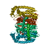
 Basic information
 Components
 Keywords
 Function and homology information
 Homo sapiens (human)
 X-RAY DIFFRACTION / NEUTRON DIFFRACTION /
  Authors
 Citation
 Structure visualization
 Downloads & links
 Other downloads
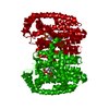


 PDBj
PDBj


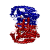
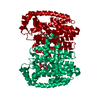
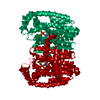
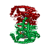
 Assembly
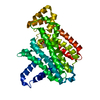


 Mass: 24.305 Da / Num. of mol.: 3 / Source method: obtained synthetically / Formula: Mg
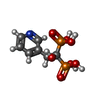
 Mass: 283.112 Da / Num. of mol.: 1 / Source method: obtained synthetically / Formula: C7H11NO7P2
 Mass: 18.015 Da / Num. of mol.: 106 / Source method: isolated from a natural source / Formula: D2O
 Sample preparation
 Processing