National Institutes of Health/National Institute Of Allergy and Infectious Diseases (NIH/NIAID)
P01 AI082362
United States
National Institutes of Health/National Institute Of Allergy and Infectious Diseases (NIH/NIAID)
UM1 AI100663
United States
National Institutes of Health/National Institute Of Allergy and Infectious Diseases (NIH/NIAID)
R01 AI084817
United States
National Institutes of Health/National Institute Of Allergy and Infectious Diseases (NIH/NIAID)
AI114401
United States
National Institutes of Health/National Institute Of Allergy and Infectious Diseases (NIH/NIAID)
AI098602
United States
Bill & Melinda Gates Foundation
United States
Citation
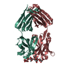
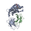
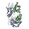
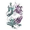
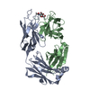
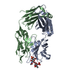
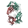
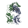
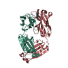
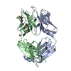
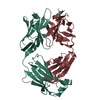
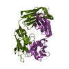
Journal: Nat Commun / Year: 2015 Title: Antibodies to a conformational epitope on gp41 neutralize HIV-1 by destabilizing the Env spike. Authors: Jeong Hyun Lee / Daniel P Leaman / Arthur S Kim / Alba Torrents de la Peña / Kwinten Sliepen / Anila Yasmeen / Ronald Derking / Alejandra Ramos / Steven W de Taeye / Gabriel Ozorowski / ...Authors: Jeong Hyun Lee / Daniel P Leaman / Arthur S Kim / Alba Torrents de la Peña / Kwinten Sliepen / Anila Yasmeen / Ronald Derking / Alejandra Ramos / Steven W de Taeye / Gabriel Ozorowski / Florian Klein / Dennis R Burton / Michel C Nussenzweig / Pascal Poignard / John P Moore / Per Johan Klasse / Rogier W Sanders / Michael B Zwick / Ian A Wilson / Andrew B Ward / Abstract: The recent identification of three broadly neutralizing antibodies (bnAbs) against gp120-gp41 interface epitopes has expanded the targetable surface on the HIV-1 envelope glycoprotein (Env) trimer. ...The recent identification of three broadly neutralizing antibodies (bnAbs) against gp120-gp41 interface epitopes has expanded the targetable surface on the HIV-1 envelope glycoprotein (Env) trimer. By using biochemical, biophysical and computational methods, we map the previously unknown trimer epitopes of two related antibodies, 3BC315 and 3BC176. A cryo-EM reconstruction of a soluble Env trimer bound to 3BC315 Fab at 9.3 Å resolution reveals that the antibody binds between two gp41 protomers, and neutralizes the virus by accelerating trimer decay. In contrast, bnAb 35O22 binding to a partially overlapping quaternary epitope at the gp120-gp41 interface does not induce decay. A conserved gp41-proximal glycan at N88 was also shown to play a role in the binding kinetics of 3BC176 and 3BC315. Finally, our data suggest that the dynamic structure of the Env trimer influences exposure of bnAb epitopes.
Mass: 24999.240 Da / Num. of mol.: 1 Source method: isolated from a genetically manipulated source Source: (gene. exp.) Homo sapiens (human) / Cell line (production host): HEK293F / Production host: Homo sapiens (human)
#2: Antibody
Antibody3BC315Fablightchain
Mass: 23009.605 Da / Num. of mol.: 1 Source method: isolated from a genetically manipulated source Source: (gene. exp.) Homo sapiens (human) / Cell line (production host): HEK293F / Production host: Homo sapiens (human)
In the structure databanks used in Yorodumi, some data are registered as the other names, "COVID-19 virus" and "2019-nCoV". Here are the details of the virus and the list of structure data.
Jan 31, 2019. EMDB accession codes are about to change! (news from PDBe EMDB page)
EMDB accession codes are about to change! (news from PDBe EMDB page)
The allocation of 4 digits for EMDB accession codes will soon come to an end. Whilst these codes will remain in use, new EMDB accession codes will include an additional digit and will expand incrementally as the available range of codes is exhausted. The current 4-digit format prefixed with “EMD-” (i.e. EMD-XXXX) will advance to a 5-digit format (i.e. EMD-XXXXX), and so on. It is currently estimated that the 4-digit codes will be depleted around Spring 2019, at which point the 5-digit format will come into force.
The EM Navigator/Yorodumi systems omit the EMD- prefix.
Related info.:Q: What is EMD? / ID/Accession-code notation in Yorodumi/EM Navigator
Yorodumi is a browser for structure data from EMDB, PDB, SASBDB, etc.
This page is also the successor to EM Navigator detail page, and also detail information page/front-end page for Omokage search.
The word "yorodu" (or yorozu) is an old Japanese word meaning "ten thousand". "mi" (miru) is to see.
Related info.:EMDB / PDB / SASBDB / Comparison of 3 databanks / Yorodumi Search / Aug 31, 2016. New EM Navigator & Yorodumi / Yorodumi Papers / Jmol/JSmol / Function and homology information / Changes in new EM Navigator and Yorodumi
 Movie
Movie Controller

 Yorodumi
 Open data
 Basic information
 Components
 Keywords
 Function and homology information
 Homo sapiens (human)
 X-RAY DIFFRACTION /
  Authors
 United States, 6items
  Citation
 Structure visualization
 Downloads & links
 Other downloads


 PDBj
PDBj


 Assembly

 Mass: 221.317 Da / Num. of mol.: 1 / Source method: obtained synthetically / Formula: C9H19NO3S / Comment: pH buffer*YM
 Mass: 18.015 Da / Num. of mol.: 339 / Source method: isolated from a natural source / Formula: H2O
 Sample preparation
 Processing