 Movie
Movie Controller

[English] 日本語
 Yorodumi
- PDB-5cb8: Crystal structure of Adenosine-5'-phosphosulfate kinase in comple... -
+ Open data

- Basic information
| Entry | Database: PDB / ID: 5cb8 | ||||||
|---|---|---|---|---|---|---|---|
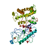
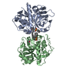
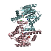
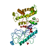
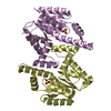
| Title | Crystal structure of Adenosine-5'-phosphosulfate kinase in complex with APS and sulfate | ||||||
 Components Components | Probable adenylyl-sulfate kinase | ||||||
 Keywords Keywords | TRANSFERASE / Synechocystis / sulfur metabolism / kinase | ||||||
| Function / homology |  Function and homology information Function and homology information: / adenylyl-sulfate kinase / adenylylsulfate kinase activity / sulfate adenylyltransferase (ATP) activity / hydrogen sulfide biosynthetic process / ATP binding Similarity search - Function | ||||||
| Biological species |  | ||||||
| Method |  X-RAY DIFFRACTION / SYNCHROTRON / MOLECULAR REPLACEMENT / Resolution: 1.88 Å X-RAY DIFFRACTION / SYNCHROTRON / MOLECULAR REPLACEMENT / Resolution: 1.88 Å | ||||||
 Authors Authors | Herrmann, J. / Jez, J.M. | ||||||
 Citation Citation | Journal: J.Biol.Chem. / Year: 2015 Title: Recapitulating the Structural Evolution of Redox Regulation in Adenosine 5'-Phosphosulfate Kinase from Cyanobacteria to Plants. Authors: Herrmann, J. / Nathin, D. / Lee, S.G. / Sun, T. / Jez, J.M. | ||||||
| History |
|
- Structure visualization
| Structure viewer | Molecule: MolmilJmol/JSmol |
|---|
- Downloads & links
-Download
| PDBx/mmCIF format | 5cb8.cif.gz | 168.2 KB | Display | PDBx/mmCIF format |
|---|---|---|---|---|
| PDB format | pdb5cb8.ent.gz | 132.1 KB | Display | PDB format |
| PDBx/mmJSON format | 5cb8.json.gz | Tree view | PDBx/mmJSON format | |
| Others |  Other downloads Other downloads |
-Validation report
| Arichive directory | https://data.pdbj.org/pub/pdb/validation_reports/cb/5cb8ftp://data.pdbj.org/pub/pdb/validation_reports/cb/5cb8 | HTTPS FTP |
|---|
-Related structure data
| Related structure data |  5cb6SC S: Starting model for refinement C: citing same article ( |
|---|---|
| Similar structure data |


-Links
 PDBj
PDBj
- Assembly
| Deposited unit | 
| ||||||||
|---|---|---|---|---|---|---|---|---|---|
| 1 |
| ||||||||
| Unit cell |
|
-Components
| #1: Protein | Mass: 21872.799 Da / Num. of mol.: 2 Source method: isolated from a genetically manipulated source Source: (gene. exp.) Strain: PCC 6803 / Kazusa / Gene: cysC, slr0676 / Production host: #2: Chemical |   Type: RNA linking / Mass: 427.284 Da / Num. of mol.: 2 / Source method: obtained synthetically / Formula: C10H14N5O10PS Type: RNA linking / Mass: 427.284 Da / Num. of mol.: 2 / Source method: obtained synthetically / Formula: C10H14N5O10PS#3: Chemical | ChemComp-SO4 /   Mass: 96.063 Da / Num. of mol.: 4 / Source method: obtained synthetically / Formula: SO4 Mass: 96.063 Da / Num. of mol.: 4 / Source method: obtained synthetically / Formula: SO4#4: Chemical |   Mass: 59.044 Da / Num. of mol.: 2 / Source method: obtained synthetically / Formula: C2H3O2 Mass: 59.044 Da / Num. of mol.: 2 / Source method: obtained synthetically / Formula: C2H3O2#5: Water | ChemComp-HOH / |  Mass: 18.015 Da / Num. of mol.: 557 / Source method: isolated from a natural source / Formula: H2O Mass: 18.015 Da / Num. of mol.: 557 / Source method: isolated from a natural source / Formula: H2O |
|---|
-Experimental details
-Experiment
| Experiment | Method: X-RAY DIFFRACTION |
|---|
- Sample preparation
| Crystal | Density Matthews: 2.95 Å3/Da / Density % sol: 58.29 % |
|---|---|
| Crystal grow | Temperature: 277 K / Method: vapor diffusion, hanging drop Details: 5 mM APS and 10 mM K2SO4 and mixed with 0.1 M CAPS/KOH, pH 10.5, 2 M ammonium sulfate, and 0.2 M LiSO4 PH range: 10.5 |
-Data collection
| Diffraction | Mean temperature: 100 K |
|---|---|
| Diffraction source | Source: SYNCHROTRON / Site: APS  / Beamline: 19-BM / Wavelength: 0.98 Å / Beamline: 19-BM / Wavelength: 0.98 Å |
| Detector | Type: SBC-3 / Detector: CCD / Date: Dec 12, 2012 |
| Radiation | Protocol: SINGLE WAVELENGTH / Monochromatic (M) / Laue (L): M / Scattering type: x-ray |
| Radiation wavelength | Wavelength: 0.98 Å / Relative weight: 1 |
| Reflection | Resolution: 1.88→31.5 Å / Num. all: 810057 / Num. obs: 37779 / % possible obs: 100 % / Redundancy: 21.4 % / Net I/σ(I): 50 |
- Processing
| Software |
| |||||||||||||||||||||||||||||||||||||||||||||||||||||||||||||||||||||||||||||||||||||||||||||||||||||||||||||||||||||||||||||||||||||||||||||||||||||||||||||||||||||||||||||||
|---|---|---|---|---|---|---|---|---|---|---|---|---|---|---|---|---|---|---|---|---|---|---|---|---|---|---|---|---|---|---|---|---|---|---|---|---|---|---|---|---|---|---|---|---|---|---|---|---|---|---|---|---|---|---|---|---|---|---|---|---|---|---|---|---|---|---|---|---|---|---|---|---|---|---|---|---|---|---|---|---|---|---|---|---|---|---|---|---|---|---|---|---|---|---|---|---|---|---|---|---|---|---|---|---|---|---|---|---|---|---|---|---|---|---|---|---|---|---|---|---|---|---|---|---|---|---|---|---|---|---|---|---|---|---|---|---|---|---|---|---|---|---|---|---|---|---|---|---|---|---|---|---|---|---|---|---|---|---|---|---|---|---|---|---|---|---|---|---|---|---|---|---|---|---|---|---|
| Refinement | Method to determine structure: MOLECULAR REPLACEMENT Starting model: 5CB6 Resolution: 1.88→31.474 Å / SU ML: 0.18 / Cross valid method: FREE R-VALUE / σ(F): 1.34 / Phase error: 18.08 / Stereochemistry target values: ML
| |||||||||||||||||||||||||||||||||||||||||||||||||||||||||||||||||||||||||||||||||||||||||||||||||||||||||||||||||||||||||||||||||||||||||||||||||||||||||||||||||||||||||||||||
| Solvent computation | Shrinkage radii: 0.9 Å / VDW probe radii: 1.11 Å / Solvent model: FLAT BULK SOLVENT MODEL | |||||||||||||||||||||||||||||||||||||||||||||||||||||||||||||||||||||||||||||||||||||||||||||||||||||||||||||||||||||||||||||||||||||||||||||||||||||||||||||||||||||||||||||||
| Refinement step | Cycle: LAST / Resolution: 1.88→31.474 Å
| |||||||||||||||||||||||||||||||||||||||||||||||||||||||||||||||||||||||||||||||||||||||||||||||||||||||||||||||||||||||||||||||||||||||||||||||||||||||||||||||||||||||||||||||
| Refine LS restraints |
| |||||||||||||||||||||||||||||||||||||||||||||||||||||||||||||||||||||||||||||||||||||||||||||||||||||||||||||||||||||||||||||||||||||||||||||||||||||||||||||||||||||||||||||||
| LS refinement shell |
| |||||||||||||||||||||||||||||||||||||||||||||||||||||||||||||||||||||||||||||||||||||||||||||||||||||||||||||||||||||||||||||||||||||||||||||||||||||||||||||||||||||||||||||||
| Refinement TLS params. | Method: refined / Refine-ID: X-RAY DIFFRACTION
| |||||||||||||||||||||||||||||||||||||||||||||||||||||||||||||||||||||||||||||||||||||||||||||||||||||||||||||||||||||||||||||||||||||||||||||||||||||||||||||||||||||||||||||||
| Refinement TLS group |
|
