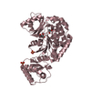
 Movie
Movie Controller


+ Open data

- Basic information
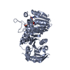
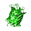
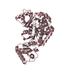
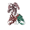
| Entry | Database: PDB / ID: 5c82 | |||||||||
|---|---|---|---|---|---|---|---|---|---|---|
| Title | Crystal structure of Nourseothricin acetyltransferase | |||||||||
 Components Components | Nourseothricin acetyltransferase | |||||||||
 Keywords Keywords | TRANSFERASE / Acettyl transferase / antibiotic ressistance / Se-Met SAD | |||||||||
| Function / homology |  Function and homology information Function and homology informationacyltransferase activity, transferring groups other than amino-acyl groups Similarity search - Function | |||||||||
| Biological species |  Streptomyces noursei (bacteria) Streptomyces noursei (bacteria) | |||||||||
| Method |  X-RAY DIFFRACTION / SYNCHROTRON / SAD / Resolution: 2.2 Å X-RAY DIFFRACTION / SYNCHROTRON / SAD / Resolution: 2.2 Å | |||||||||
 Authors Authors | Kumar, D. / Ghosh, A. / Taneja, B. / Chakraborty, K. | |||||||||
| Funding support |  India, 1items India, 1items
| |||||||||
 Citation Citation | Journal: To Be Published Title: Crystal structure of Nourseothricin acetyltransferase Authors: Kumar, D. / Ghosh, A. / Taneja, B. / Chakraborty, K. | |||||||||
| History |
|
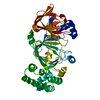
- Structure visualization
| Structure viewer | Molecule: MolmilJmol/JSmol |
|---|
- Downloads & links
-Download
| PDBx/mmCIF format | 5c82.cif.gz | 50.2 KB | Display | PDBx/mmCIF format |
|---|---|---|---|---|
| PDB format | pdb5c82.ent.gz | 34.1 KB | Display | PDB format |
| PDBx/mmJSON format | 5c82.json.gz | Tree view | PDBx/mmJSON format | |
| Others |  Other downloads Other downloads |
-Validation report
| Arichive directory | https://data.pdbj.org/pub/pdb/validation_reports/c8/5c82ftp://data.pdbj.org/pub/pdb/validation_reports/c8/5c82 | HTTPS FTP |
|---|
-Related structure data
| Similar structure data |
|---|


-Links
 PDBj
PDBj


- Assembly
| Deposited unit | 
| ||||||||
|---|---|---|---|---|---|---|---|---|---|
| 1 | 
| ||||||||
| Unit cell |
|
-Components
| #1: Protein | Mass: 20790.867 Da / Num. of mol.: 1 Source method: isolated from a genetically manipulated source Source: (gene. exp.) Streptomyces noursei (bacteria) / Gene: nat1 / Plasmid: pET-SUMO / Production host: |
|---|---|
| #2: Chemical | ChemComp-TAR /   Mass: 150.087 Da / Num. of mol.: 1 / Source method: obtained synthetically / Formula: C4H6O6 Mass: 150.087 Da / Num. of mol.: 1 / Source method: obtained synthetically / Formula: C4H6O6 |
| #3: Water | ChemComp-HOH /  Mass: 18.015 Da / Num. of mol.: 97 / Source method: isolated from a natural source / Formula: H2O Mass: 18.015 Da / Num. of mol.: 97 / Source method: isolated from a natural source / Formula: H2O |
| Has protein modification | Y |
-Experimental details
-Experiment
| Experiment | Method: X-RAY DIFFRACTION |
|---|
- Sample preparation
| Crystal | Density Matthews: 2.11 Å3/Da / Density % sol: 41.75 % Description: THE ENTRY CONTAINS FRIEDEL PAIRS IN F_PLUS/MINUS COLUMNS. |
|---|---|
| Crystal grow | Temperature: 297.15 K / Method: vapor diffusion, sitting drop / pH: 8.5 Details: 0.1 M Carboxylic acids (0.2M Sodium formate; 0.2M Ammonium acetate; 0.2M Sodium citrate tribasic dihydrate; 0.2M Sodium potassium tartrate tetrahydrate; 0.2M Sodium oxamate), 0.1 M Buffer ...Details: 0.1 M Carboxylic acids (0.2M Sodium formate; 0.2M Ammonium acetate; 0.2M Sodium citrate tribasic dihydrate; 0.2M Sodium potassium tartrate tetrahydrate; 0.2M Sodium oxamate), 0.1 M Buffer System 3(Tris base; BICINE) 8.5, 50 % v/v Precipitant Mix 4(25% v/v MPD; 25% PEG 1000; 25% w/v PEG 3350) |
-Data collection
| Diffraction | Mean temperature: 100 K |
|---|---|
| Diffraction source | Source: SYNCHROTRON / Site: RRCAT INDUS-2 / Beamline: PX-BL21 / Wavelength: 0.97869 Å |
| Detector | Type: RAYONIX MX-225 / Detector: CCD / Date: Jun 9, 2015 |
| Radiation | Monochromator: Si (111) and Si (220) Double crystal monochromato Protocol: SINGLE WAVELENGTH / Monochromatic (M) / Laue (L): M / Scattering type: x-ray |
| Radiation wavelength | Wavelength: 0.97869 Å / Relative weight: 1 |
| Reflection | Resolution: 2.2→33.82 Å / Num. obs: 9075 / % possible obs: 98.7 % / Redundancy: 7.2 % / Biso Wilson estimate: 29.45 Å2 / Rmerge(I) obs: 0.091 / Net I/σ(I): 21.4 |
| Reflection shell | Resolution: 2.2→2.27 Å / Redundancy: 5.1 % / Rmerge(I) obs: 0.341 / % possible all: 90.8 |
- Processing
| Software |
| ||||||||||||||||||||||||||||||||||||||||||||||||||||||||||||||||||||||||||||||||||||||||||||||||||||||||||||||||||
|---|---|---|---|---|---|---|---|---|---|---|---|---|---|---|---|---|---|---|---|---|---|---|---|---|---|---|---|---|---|---|---|---|---|---|---|---|---|---|---|---|---|---|---|---|---|---|---|---|---|---|---|---|---|---|---|---|---|---|---|---|---|---|---|---|---|---|---|---|---|---|---|---|---|---|---|---|---|---|---|---|---|---|---|---|---|---|---|---|---|---|---|---|---|---|---|---|---|---|---|---|---|---|---|---|---|---|---|---|---|---|---|---|---|---|---|
| Refinement | Method to determine structure: SAD / Resolution: 2.2→15.28 Å / Cor.coef. Fo:Fc: 0.9328 / Cor.coef. Fo:Fc free: 0.9031 / SU R Cruickshank DPI: 0.257 / Cross valid method: THROUGHOUT / σ(F): 0 / SU R Blow DPI: 0.269 / SU Rfree Blow DPI: 0.196 / SU Rfree Cruickshank DPI: 0.195 Details: SF FILE CONTAINS FRIEDEL PAIRS UNDER I/F_MINUS AND I/F_PLUS COLUMNS.
| ||||||||||||||||||||||||||||||||||||||||||||||||||||||||||||||||||||||||||||||||||||||||||||||||||||||||||||||||||
| Displacement parameters | Biso mean: 29.43 Å2
| ||||||||||||||||||||||||||||||||||||||||||||||||||||||||||||||||||||||||||||||||||||||||||||||||||||||||||||||||||
| Refine analyze | Luzzati coordinate error obs: 0.269 Å | ||||||||||||||||||||||||||||||||||||||||||||||||||||||||||||||||||||||||||||||||||||||||||||||||||||||||||||||||||
| Refinement step | Cycle: 1 / Resolution: 2.2→15.28 Å
| ||||||||||||||||||||||||||||||||||||||||||||||||||||||||||||||||||||||||||||||||||||||||||||||||||||||||||||||||||
| Refine LS restraints |
| ||||||||||||||||||||||||||||||||||||||||||||||||||||||||||||||||||||||||||||||||||||||||||||||||||||||||||||||||||
| LS refinement shell | Resolution: 2.2→2.46 Å / Total num. of bins used: 5
|
