 Movie
Movie Controller

[English] 日本語
 Yorodumi
- PDB-5c80: X-ray structure uridine phosphorylase from Vibrio cholerae in com... -
+ Open data

- Basic information
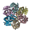
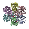
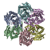
| Entry | Database: PDB / ID: 5c80 | ||||||
|---|---|---|---|---|---|---|---|
| Title | X-ray structure uridine phosphorylase from Vibrio cholerae in complex with uridine at 2.24 A resolution | ||||||
 Components Components | Uridine phosphorylase | ||||||
 Keywords Keywords | TRANSFERASE / Rossmann Fold | ||||||
| Function / homology |  Function and homology information Function and homology informationnucleotide catabolic process / uridine phosphorylase / nucleoside catabolic process / UMP salvage / uridine phosphorylase activity / metal ion binding / cytosol Similarity search - Function | ||||||
| Biological species |   Vibrio cholerae (bacteria) Vibrio cholerae (bacteria) | ||||||
| Method |  X-RAY DIFFRACTION / SYNCHROTRON / MOLECULAR REPLACEMENT / molecular replacement / Resolution: 2.243 Å X-RAY DIFFRACTION / SYNCHROTRON / MOLECULAR REPLACEMENT / molecular replacement / Resolution: 2.243 Å | ||||||
 Authors Authors | Prokofev, I.I. / Lashkov, A.A. / Gabdoulkhakov, A.G. / Betzel, C. / Mikhailov, A.M. | ||||||
| Funding support |  Russian Federation, 1items Russian Federation, 1items
| ||||||
 Citation Citation | Journal: Crystallography Reports / Year: 2016 Title: X-ray structures of uridine phosphorylase from Vibrio cholerae in complexes with uridine, thymidine, uracil, thymine, and phosphate anion: Substrate specificity of bacterial uridine phosphorylases Authors: Prokofev, I.I. / Lashkov, A.A. / Gabdoulkhakov, A.G. / Balaev, V.V. / Seregina, T.A. / Mironov, A.S. / Betzel, C. / Mikhailov, A.M. | ||||||
| History |
|
- Structure visualization
| Structure viewer | Molecule: MolmilJmol/JSmol |
|---|
- Downloads & links
-Download
| PDBx/mmCIF format | 5c80.cif.gz | 313.3 KB | Display | PDBx/mmCIF format |
|---|---|---|---|---|
| PDB format | pdb5c80.ent.gz | 252 KB | Display | PDB format |
| PDBx/mmJSON format | 5c80.json.gz | Tree view | PDBx/mmJSON format | |
| Others |  Other downloads Other downloads |
-Validation report
| Arichive directory | https://data.pdbj.org/pub/pdb/validation_reports/c8/5c80ftp://data.pdbj.org/pub/pdb/validation_reports/c8/5c80 | HTTPS FTP |
|---|
-Related structure data
| Related structure data |  4ip0C  4lzwC  4lwzS S: Starting model for refinement C: citing same article ( |
|---|---|
| Similar structure data |


-Links
 PDBj
PDBj- Assembly
| Deposited unit | 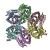
| ||||||||
|---|---|---|---|---|---|---|---|---|---|
| 1 |
| ||||||||
| Unit cell |
|
-Components
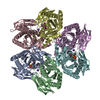
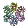
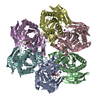
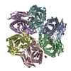
-Protein , 1 types, 6 molecules ABCDEF
| #1: Protein | Mass: 27108.994 Da / Num. of mol.: 6 Source method: isolated from a genetically manipulated source Source: (gene. exp.) Vibrio cholerae (bacteria)Gene: udp, DN30_1909, VC39_02535, VC78_02550, VS27_10630, WG08_05660 Production host: |
|---|
-Non-polymers , 7 types, 851 molecules 


| #2: Chemical | ChemComp-URI /  Mass: 244.201 Da / Num. of mol.: 6 / Source method: obtained synthetically / Formula: C9H12N2O6 Mass: 244.201 Da / Num. of mol.: 6 / Source method: obtained synthetically / Formula: C9H12N2O6#3: Chemical | ChemComp-TRS /  Mass: 122.143 Da / Num. of mol.: 9 / Source method: obtained synthetically / Formula: C4H12NO3 / Comment: pH buffer*YM Mass: 122.143 Da / Num. of mol.: 9 / Source method: obtained synthetically / Formula: C4H12NO3 / Comment: pH buffer*YM#4: Chemical |  Mass: 106.120 Da / Num. of mol.: 3 / Source method: obtained synthetically / Formula: C4H10O3 Mass: 106.120 Da / Num. of mol.: 3 / Source method: obtained synthetically / Formula: C4H10O3#5: Chemical | ChemComp-CL /  Mass: 35.453 Da / Num. of mol.: 6 / Source method: obtained synthetically / Formula: Cl Mass: 35.453 Da / Num. of mol.: 6 / Source method: obtained synthetically / Formula: Cl#6: Chemical |  Mass: 92.094 Da / Num. of mol.: 3 / Source method: obtained synthetically / Formula: C3H8O3 Mass: 92.094 Da / Num. of mol.: 3 / Source method: obtained synthetically / Formula: C3H8O3#7: Chemical |  Mass: 22.990 Da / Num. of mol.: 2 / Source method: obtained synthetically / Formula: Na Mass: 22.990 Da / Num. of mol.: 2 / Source method: obtained synthetically / Formula: Na#8: Water | ChemComp-HOH / | Mass: 18.015 Da / Num. of mol.: 822 / Source method: isolated from a natural source / Formula: H2O |
|---|
-Experimental details
-Experiment
| Experiment | Method: X-RAY DIFFRACTION / Number of used crystals: 1 |
|---|
- Sample preparation
| Crystal | Density Matthews: 2.12 Å3/Da / Density % sol: 41.99 % |
|---|---|
| Crystal grow | Temperature: 293 K / Method: vapor diffusion, hanging drop / pH: 8.5 / Details: PEG4000, 0.1M TRIS-HCl, 0.2M MgCl2x6H2O |
-Data collection
| Diffraction | Mean temperature: 100 K | ||||||||||||||||||||||||||||||||||||||||||||||||||||||||||||||||||||||||||||||||||||||||||||||||||||||||||||||
|---|---|---|---|---|---|---|---|---|---|---|---|---|---|---|---|---|---|---|---|---|---|---|---|---|---|---|---|---|---|---|---|---|---|---|---|---|---|---|---|---|---|---|---|---|---|---|---|---|---|---|---|---|---|---|---|---|---|---|---|---|---|---|---|---|---|---|---|---|---|---|---|---|---|---|---|---|---|---|---|---|---|---|---|---|---|---|---|---|---|---|---|---|---|---|---|---|---|---|---|---|---|---|---|---|---|---|---|---|---|---|---|
| Diffraction source | Source: SYNCHROTRON / Site: BESSY  / Beamline: 14.1 / Wavelength: 0.97989 Å / Beamline: 14.1 / Wavelength: 0.97989 Å | ||||||||||||||||||||||||||||||||||||||||||||||||||||||||||||||||||||||||||||||||||||||||||||||||||||||||||||||
| Detector | Type: PSI PILATUS 6M / Detector: PIXEL / Date: May 16, 2015 | ||||||||||||||||||||||||||||||||||||||||||||||||||||||||||||||||||||||||||||||||||||||||||||||||||||||||||||||
| Radiation | Protocol: SINGLE WAVELENGTH / Monochromatic (M) / Laue (L): M / Scattering type: x-ray | ||||||||||||||||||||||||||||||||||||||||||||||||||||||||||||||||||||||||||||||||||||||||||||||||||||||||||||||
| Radiation wavelength | Wavelength: 0.97989 Å / Relative weight: 1 | ||||||||||||||||||||||||||||||||||||||||||||||||||||||||||||||||||||||||||||||||||||||||||||||||||||||||||||||
| Reflection | Resolution: 2.243→77.231 Å / Num. all: 58293 / Num. obs: 58293 / % possible obs: 91.1 % / Redundancy: 1.7 % / Rmerge(I) obs: 0.055 / Rpim(I) all: 0.055 / Rrim(I) all: 0.077 / Net I/av σ(I): 11.229 / Net I/σ(I): 9.6 / Num. measured all: 101646 | ||||||||||||||||||||||||||||||||||||||||||||||||||||||||||||||||||||||||||||||||||||||||||||||||||||||||||||||
| Reflection shell | Diffraction-ID: 1 / Rejects: _
|
-Phasing
| Phasing | Method: molecular replacement | |||||||||
|---|---|---|---|---|---|---|---|---|---|---|
| Phasing MR | Model details: Phaser MODE: MR_AUTO
|
- Processing
| Software |
| ||||||||||||||||||||||||||||||||||||||||||||||||||||||||||||
|---|---|---|---|---|---|---|---|---|---|---|---|---|---|---|---|---|---|---|---|---|---|---|---|---|---|---|---|---|---|---|---|---|---|---|---|---|---|---|---|---|---|---|---|---|---|---|---|---|---|---|---|---|---|---|---|---|---|---|---|---|---|
| Refinement | Method to determine structure: MOLECULAR REPLACEMENT Starting model: 4LWZ Resolution: 2.243→45.47 Å / Cor.coef. Fo:Fc: 0.957 / Cor.coef. Fo:Fc free: 0.918 / WRfactor Rfree: 0.2136 / WRfactor Rwork: 0.1515 / FOM work R set: 0.8352 / SU B: 7.17 / SU ML: 0.174 / SU R Cruickshank DPI: 0.5856 / SU Rfree: 0.2644 / Cross valid method: THROUGHOUT / σ(F): 0 / ESU R: 0.586 / ESU R Free: 0.264 / Stereochemistry target values: MAXIMUM LIKELIHOOD / Details: U VALUES : REFINED INDIVIDUALLY
| ||||||||||||||||||||||||||||||||||||||||||||||||||||||||||||
| Solvent computation | Ion probe radii: 0.8 Å / Shrinkage radii: 0.8 Å / VDW probe radii: 1.2 Å / Solvent model: MASK | ||||||||||||||||||||||||||||||||||||||||||||||||||||||||||||
| Displacement parameters | Biso max: 103.62 Å2 / Biso mean: 30.999 Å2 / Biso min: 3.04 Å2
| ||||||||||||||||||||||||||||||||||||||||||||||||||||||||||||
| Refinement step | Cycle: final / Resolution: 2.243→45.47 Å
| ||||||||||||||||||||||||||||||||||||||||||||||||||||||||||||
| Refine LS restraints |
| ||||||||||||||||||||||||||||||||||||||||||||||||||||||||||||
| LS refinement shell | Resolution: 2.243→2.301 Å / Total num. of bins used: 20
|
