 Movie
Movie Controller

[English] 日本語
 Yorodumi
- PDB-5c4q: Crystal Structure Analysis of bromodomain from Leishmania donovan... -
+ Open data

- Basic information
| Entry | Database: PDB / ID: 5c4q | ||||||
|---|---|---|---|---|---|---|---|
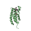
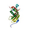
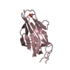
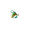
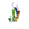
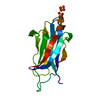
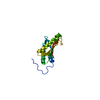
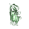
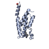
| Title | Crystal Structure Analysis of bromodomain from Leishmania donovani complexed with bromosporine | ||||||
 Components Components | Bromodomain | ||||||
 Keywords Keywords | TRANSCRIPTION / Structural Genomics Consortium / SGC / Bromodomain / Bromosporine / Leishmania donovani | ||||||
| Function / homology |  Function and homology information Function and homology information: / Bromodomain-like / Histone Acetyltransferase; Chain A / Bromodomain / bromo domain / Bromodomain / Bromodomain (BrD) profile. / Bromodomain-like superfamily / Up-down Bundle / Mainly Alpha Similarity search - Domain/homology | ||||||
| Biological species |  Leishmania donovani (eukaryote) Leishmania donovani (eukaryote) | ||||||
| Method |  X-RAY DIFFRACTION / MOLECULAR REPLACEMENT / Resolution: 1.932 Å X-RAY DIFFRACTION / MOLECULAR REPLACEMENT / Resolution: 1.932 Å | ||||||
 Authors Authors | Jiang, D.Q. / Tempel, W. / Loppnau, P. / Graslund, S. / Arrowsmith, C.H. / Edwards, A.M. / Bountra, C. / Hui, R. / Amani, M. / Hou, C.F.D. / Structural Genomics Consortium (SGC) | ||||||
 Citation Citation | Journal: to be published Title: Crystal Structure Analysis of bromodomain from Leishmania donovani complexed with bromosporine Authors: Jiang, D.Q. / Tempel, W. / Loppnau, P. / Graslund, S. / Arrowsmith, C.H. / Edwards, A.M. / Bountra, C. / Hui, R. / Amani, M. / Hou, C.F.D. | ||||||
| History |
|
- Structure visualization
| Structure viewer | Molecule: MolmilJmol/JSmol |
|---|
- Downloads & links
-Download
| PDBx/mmCIF format | 5c4q.cif.gz | 64.5 KB | Display | PDBx/mmCIF format |
|---|---|---|---|---|
| PDB format | pdb5c4q.ent.gz | 46.6 KB | Display | PDB format |
| PDBx/mmJSON format | 5c4q.json.gz | Tree view | PDBx/mmJSON format | |
| Others |  Other downloads Other downloads |
-Validation report
| Arichive directory | https://data.pdbj.org/pub/pdb/validation_reports/c4/5c4qftp://data.pdbj.org/pub/pdb/validation_reports/c4/5c4q | HTTPS FTP |
|---|
-Related structure data
| Related structure data | 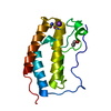 4hbwS S: Starting model for refinement |
|---|---|
| Similar structure data |

-Links
 PDBj
PDBj- Assembly
| Deposited unit | 
| ||||||||
|---|---|---|---|---|---|---|---|---|---|
| 1 | 
| ||||||||
| 2 | 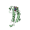
| ||||||||
| 3 |
| ||||||||
| Unit cell |
| ||||||||
| Details | The biological assembly of the protein indicated by Mass spectrometry and Gel filtration is monomer. |
-Components
| #1: Protein | Mass: 14440.311 Da / Num. of mol.: 2 Source method: isolated from a genetically manipulated source Source: (gene. exp.) Leishmania donovani (strain BPK282A1) (eukaryote)Strain: BPK282A1 / Gene: LDBPK_363130 / Plasmid: V3R pRARE2 / Production host:  #2: Chemical |   Mass: 404.444 Da / Num. of mol.: 2 / Source method: obtained synthetically / Formula: C17H20N6O4S Mass: 404.444 Da / Num. of mol.: 2 / Source method: obtained synthetically / Formula: C17H20N6O4S#3: Chemical | ChemComp-UNX /   Num. of mol.: 9 / Source method: obtained synthetically Num. of mol.: 9 / Source method: obtained synthetically#4: Water | ChemComp-HOH / |  Mass: 18.015 Da / Num. of mol.: 140 / Source method: isolated from a natural source / Formula: H2O Mass: 18.015 Da / Num. of mol.: 140 / Source method: isolated from a natural source / Formula: H2O |
|---|
-Experimental details
-Experiment
| Experiment | Method: X-RAY DIFFRACTION / Number of used crystals: 1 |
|---|
- Sample preparation
| Crystal | Density Matthews: 2.03 Å3/Da / Density % sol: 39.36 % |
|---|---|
| Crystal grow | Temperature: 293 K / Method: vapor diffusion, sitting drop / pH: 7 / Details: 2.4 M sodium malonate |
-Data collection
| Diffraction | Mean temperature: 100 K | |||||||||||||||||||||||||||||||||||||||||||||||||||||||||||||||||||||||||||||||||||||||||||||||||||||||||||||||||||||||||||||||||||||||||||||||||||||||||||||||||||||||||||||||||||||||||||||
|---|---|---|---|---|---|---|---|---|---|---|---|---|---|---|---|---|---|---|---|---|---|---|---|---|---|---|---|---|---|---|---|---|---|---|---|---|---|---|---|---|---|---|---|---|---|---|---|---|---|---|---|---|---|---|---|---|---|---|---|---|---|---|---|---|---|---|---|---|---|---|---|---|---|---|---|---|---|---|---|---|---|---|---|---|---|---|---|---|---|---|---|---|---|---|---|---|---|---|---|---|---|---|---|---|---|---|---|---|---|---|---|---|---|---|---|---|---|---|---|---|---|---|---|---|---|---|---|---|---|---|---|---|---|---|---|---|---|---|---|---|---|---|---|---|---|---|---|---|---|---|---|---|---|---|---|---|---|---|---|---|---|---|---|---|---|---|---|---|---|---|---|---|---|---|---|---|---|---|---|---|---|---|---|---|---|---|---|---|---|---|
| Diffraction source | Source: ROTATING ANODE / Type: RIGAKU FR-E+ SUPERBRIGHT / Wavelength: 1.5418 Å | |||||||||||||||||||||||||||||||||||||||||||||||||||||||||||||||||||||||||||||||||||||||||||||||||||||||||||||||||||||||||||||||||||||||||||||||||||||||||||||||||||||||||||||||||||||||||||||
| Detector | Type: RIGAKU RAXIS IV / Detector: IMAGE PLATE / Date: Jun 9, 2015 | |||||||||||||||||||||||||||||||||||||||||||||||||||||||||||||||||||||||||||||||||||||||||||||||||||||||||||||||||||||||||||||||||||||||||||||||||||||||||||||||||||||||||||||||||||||||||||||
| Radiation | Protocol: SINGLE WAVELENGTH / Monochromatic (M) / Laue (L): M / Scattering type: x-ray | |||||||||||||||||||||||||||||||||||||||||||||||||||||||||||||||||||||||||||||||||||||||||||||||||||||||||||||||||||||||||||||||||||||||||||||||||||||||||||||||||||||||||||||||||||||||||||||
| Radiation wavelength | Wavelength: 1.5418 Å / Relative weight: 1 | |||||||||||||||||||||||||||||||||||||||||||||||||||||||||||||||||||||||||||||||||||||||||||||||||||||||||||||||||||||||||||||||||||||||||||||||||||||||||||||||||||||||||||||||||||||||||||||
| Reflection | Resolution: 1.93→20 Å / Num. obs: 16741 / % possible obs: 94.9 % / Redundancy: 3.7 % / Biso Wilson estimate: 23.97 Å2 / Rmerge(I) obs: 0.094 / Rpim(I) all: 0.055 / Rrim(I) all: 0.109 / Χ2: 1.524 / Net I/av σ(I): 14.418 / Net I/σ(I): 7.2 / Num. measured all: 61331 | |||||||||||||||||||||||||||||||||||||||||||||||||||||||||||||||||||||||||||||||||||||||||||||||||||||||||||||||||||||||||||||||||||||||||||||||||||||||||||||||||||||||||||||||||||||||||||||
| Reflection shell | Diffraction-ID: 1 / Rejects: _
|
- Processing
| Software |
| |||||||||||||||||||||||||||||||||||||||||||||||||
|---|---|---|---|---|---|---|---|---|---|---|---|---|---|---|---|---|---|---|---|---|---|---|---|---|---|---|---|---|---|---|---|---|---|---|---|---|---|---|---|---|---|---|---|---|---|---|---|---|---|---|
| Refinement | Method to determine structure: MOLECULAR REPLACEMENT Starting model: 4HBW Resolution: 1.932→19.16 Å / SU ML: 0.27 / Cross valid method: FREE R-VALUE / σ(F): 1.34 / Phase error: 25.06 / Stereochemistry target values: ML Details: Bromosporine restraints generated by PHENIX.ELBOW were modified to restrain coordinates of the lactamate moiety in a single plane, prompted by a MOGUL query of related structures in the ...Details: Bromosporine restraints generated by PHENIX.ELBOW were modified to restrain coordinates of the lactamate moiety in a single plane, prompted by a MOGUL query of related structures in the Cambridge Structural Database.
| |||||||||||||||||||||||||||||||||||||||||||||||||
| Solvent computation | Shrinkage radii: 0.9 Å / VDW probe radii: 1.11 Å / Solvent model: FLAT BULK SOLVENT MODEL | |||||||||||||||||||||||||||||||||||||||||||||||||
| Displacement parameters | Biso max: 75.05 Å2 / Biso mean: 23.9427 Å2 / Biso min: 10.67 Å2 | |||||||||||||||||||||||||||||||||||||||||||||||||
| Refinement step | Cycle: final / Resolution: 1.932→19.16 Å
| |||||||||||||||||||||||||||||||||||||||||||||||||
| Refine LS restraints |
| |||||||||||||||||||||||||||||||||||||||||||||||||
| LS refinement shell | Refine-ID: X-RAY DIFFRACTION / Total num. of bins used: 6
|
