 Movie
Movie Controller

[English] 日本語
 Yorodumi
- PDB-5bw7: Crystal structure of nonfucosylated Fc Y296W mutant complexed wit... -
+ Open data

- Basic information
| Entry | Database: PDB / ID: 5bw7 | |||||||||||||||
|---|---|---|---|---|---|---|---|---|---|---|---|---|---|---|---|---|
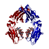
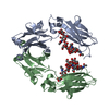
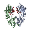
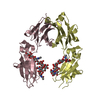
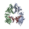
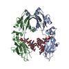
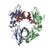
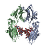
| Title | Crystal structure of nonfucosylated Fc Y296W mutant complexed with bis-glycosylated soluble form of Fc gamma receptor IIIa | |||||||||||||||
 Components Components |
| |||||||||||||||
 Keywords Keywords | IMMUNE SYSTEM / COMPLEX / FC FRAGMENT / IGG / RECEPTOR / CD16 / GAMMA | |||||||||||||||
| Function / homology |  Function and homology information Function and homology informationlow-affinity IgG receptor activity / immune receptor activity / natural killer cell degranulation / IgG receptor activity / Fc-gamma receptor III complex / Fc-gamma receptor signaling pathway / macrophage activation / complement-dependent cytotoxicity / antibody-dependent cellular cytotoxicity / Fc-gamma receptor I complex binding ...low-affinity IgG receptor activity / immune receptor activity / natural killer cell degranulation / IgG receptor activity / Fc-gamma receptor III complex / Fc-gamma receptor signaling pathway / macrophage activation / complement-dependent cytotoxicity / antibody-dependent cellular cytotoxicity / Fc-gamma receptor I complex binding / natural killer cell activation / IgG binding / immunoglobulin complex, circulating / Classical antibody-mediated complement activation / immunoglobulin receptor binding / Initial triggering of complement / IgG immunoglobulin complex / natural killer cell mediated cytotoxicity / positive regulation of natural killer cell proliferation / FCGR activation / complement activation, classical pathway / Role of phospholipids in phagocytosis / antigen binding / FCGR3A-mediated IL10 synthesis / Regulation of Complement cascade / B cell receptor signaling pathway / FCGR3A-mediated phagocytosis / phosphatidylinositol 3-kinase/protein kinase B signal transduction / calcium-mediated signaling / Regulation of actin dynamics for phagocytic cup formation / Immunoregulatory interactions between a Lymphoid and a non-Lymphoid cell / positive regulation of tumor necrosis factor production / antibacterial humoral response / Interleukin-4 and Interleukin-13 signaling / blood microparticle / adaptive immune response / cell surface receptor signaling pathway / immune response / external side of plasma membrane / : / extracellular exosome / extracellular region / plasma membrane Similarity search - Function | |||||||||||||||
| Biological species |  Homo sapiens (human) Homo sapiens (human) | |||||||||||||||
| Method |  X-RAY DIFFRACTION / SYNCHROTRON / MOLECULAR REPLACEMENT / Resolution: 3 Å X-RAY DIFFRACTION / SYNCHROTRON / MOLECULAR REPLACEMENT / Resolution: 3 Å | |||||||||||||||
 Authors Authors | Isoda, Y. / Yagi, H. / Satoh, T. / Shibata-Koyama, M. / Masuda, K. / Satoh, M. / Kato, K. / Iida, S. | |||||||||||||||
| Funding support |  Japan, 4items Japan, 4items
| |||||||||||||||
 Citation Citation | Journal: Plos One / Year: 2015 Title: Importance of the Side Chain at Position 296 of Antibody Fc in Interactions with Fc gamma RIIIa and Other Fc gamma Receptors Authors: Isoda, Y. / Yagi, H. / Satoh, T. / Shibata-Koyama, M. / Masuda, K. / Satoh, M. / Kato, K. / Iida, S. | |||||||||||||||
| History |
|
- Structure visualization
| Structure viewer | Molecule: MolmilJmol/JSmol |
|---|
- Downloads & links
-Download
| PDBx/mmCIF format | 5bw7.cif.gz | 142.6 KB | Display | PDBx/mmCIF format |
|---|---|---|---|---|
| PDB format | pdb5bw7.ent.gz | 110.2 KB | Display | PDB format |
| PDBx/mmJSON format | 5bw7.json.gz | Tree view | PDBx/mmJSON format | |
| Others |  Other downloads Other downloads |
-Validation report
| Arichive directory | https://data.pdbj.org/pub/pdb/validation_reports/bw/5bw7ftp://data.pdbj.org/pub/pdb/validation_reports/bw/5bw7 | HTTPS FTP |
|---|
-Related structure data
| Related structure data |  3ay4S S: Starting model for refinement |
|---|---|
| Similar structure data |


-Links
 PDBj
PDBj


- Assembly
| Deposited unit | 
| ||||||||
|---|---|---|---|---|---|---|---|---|---|
| 1 | 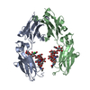
| ||||||||
| 2 | 
| ||||||||
| Unit cell |
|
-Components
-Protein , 2 types, 3 molecules ABC
| #1: Protein | Mass: 25120.469 Da / Num. of mol.: 2 / Fragment: UNP residues 108-330 / Mutation: Y296W Source method: isolated from a genetically manipulated source Source: (gene. exp.) Homo sapiens (human) / Gene: IGHG1 / Plasmid: PKANTEX2160 / Production host:   Cricetulus griseus (Chinese hamster) / Strain (production host): MS704 / References: UniProt: P01857 Cricetulus griseus (Chinese hamster) / Strain (production host): MS704 / References: UniProt: P01857#2: Protein | | Mass: 20641.012 Da / Num. of mol.: 1 / Fragment: UNP residues 21-193 / Mutation: N38Q, N74Q, F158V, N169Q Source method: isolated from a genetically manipulated source Source: (gene. exp.) Homo sapiens (human) / Gene: FCGR3A, CD16A, FCG3, FCGR3, IGFR3 / Plasmid: PKANTEX93 / Production host: Cricetulus griseus (Chinese hamster) / References: UniProt: P08637 |
|---|
-Sugars , 4 types, 4 molecules
| #3: Polysaccharide | beta-D-galactopyranose-(1-4)-2-acetamido-2-deoxy-beta-D-glucopyranose-(1-2)-alpha-D-mannopyranose- ...beta-D-galactopyranose-(1-4)-2-acetamido-2-deoxy-beta-D-glucopyranose-(1-2)-alpha-D-mannopyranose-(1-6)-[2-acetamido-2-deoxy-beta-D-glucopyranose-(1-2)-alpha-D-mannopyranose-(1-3)]beta-D-mannopyranose-(1-4)-2-acetamido-2-deoxy-beta-D-glucopyranose-(1-4)-2-acetamido-2-deoxy-beta-D-glucopyranose Source method: isolated from a genetically manipulated source |
|---|---|
| #4: Polysaccharide | 2-acetamido-2-deoxy-beta-D-glucopyranose-(1-2)-alpha-D-mannopyranose-(1-3)-[2-acetamido-2-deoxy- ...2-acetamido-2-deoxy-beta-D-glucopyranose-(1-2)-alpha-D-mannopyranose-(1-3)-[2-acetamido-2-deoxy-beta-D-glucopyranose-(1-2)-alpha-D-mannopyranose-(1-6)]beta-D-mannopyranose-(1-4)-2-acetamido-2-deoxy-beta-D-glucopyranose-(1-4)-2-acetamido-2-deoxy-beta-D-glucopyranose Source method: isolated from a genetically manipulated source |
| #5: Polysaccharide | 2-acetamido-2-deoxy-beta-D-glucopyranose-(1-2)-alpha-D-mannopyranose-(1-6)-[alpha-D-mannopyranose- ...2-acetamido-2-deoxy-beta-D-glucopyranose-(1-2)-alpha-D-mannopyranose-(1-6)-[alpha-D-mannopyranose-(1-3)]beta-D-mannopyranose-(1-4)-2-acetamido-2-deoxy-beta-D-glucopyranose-(1-4)-[alpha-L-fucopyranose-(1-6)]2-acetamido-2-deoxy-beta-D-glucopyranose Source method: isolated from a genetically manipulated source |
| #6: Polysaccharide | 2-acetamido-2-deoxy-beta-D-glucopyranose-(1-2)-alpha-D-mannopyranose-(1-3)-beta-D-mannopyranose-(1- ...2-acetamido-2-deoxy-beta-D-glucopyranose-(1-2)-alpha-D-mannopyranose-(1-3)-beta-D-mannopyranose-(1-4)-2-acetamido-2-deoxy-beta-D-glucopyranose-(1-4)-2-acetamido-2-deoxy-beta-D-glucopyranose Source method: isolated from a genetically manipulated source |
-Non-polymers , 2 types, 27 molecules 

| #7: Chemical | ChemComp-CL /  Mass: 35.453 Da / Num. of mol.: 1 / Source method: obtained synthetically / Formula: Cl Mass: 35.453 Da / Num. of mol.: 1 / Source method: obtained synthetically / Formula: Cl |
|---|---|
| #8: Water | ChemComp-HOH / Mass: 18.015 Da / Num. of mol.: 26 / Source method: isolated from a natural source / Formula: H2O |
-Details
| Has protein modification | Y |
|---|
-Experimental details
-Experiment
| Experiment | Method: X-RAY DIFFRACTION |
|---|
- Sample preparation
| Crystal | Density Matthews: 3.7 Å3/Da / Density % sol: 66.72 % |
|---|---|
| Crystal grow | Temperature: 293 K / Method: vapor diffusion, sitting drop / pH: 6.5 Details: 12% PEG20,000 0.1 M MES (pH 6.5) 1% Zwittergent 3-08 |
-Data collection
| Diffraction | Mean temperature: 95 K |
|---|---|
| Diffraction source | Source: SYNCHROTRON / Site: Photon Factory / Beamline: AR-NW12A / Wavelength: 1 Å |
| Detector | Type: ADSC QUANTUM 210r / Detector: CCD / Date: Apr 19, 2013 |
| Radiation | Protocol: SINGLE WAVELENGTH / Monochromatic (M) / Laue (L): M / Scattering type: x-ray |
| Radiation wavelength | Wavelength: 1 Å / Relative weight: 1 |
| Reflection | Resolution: 2.9→50 Å / Num. obs: 24423 / % possible obs: 98.4 % / Redundancy: 6.7 % / Biso Wilson estimate: 58.73 Å2 / Rmerge(I) obs: 0.092 / Net I/σ(I): 30.8 |
| Reflection shell | Resolution: 2.9→2.95 Å / Redundancy: 7.1 % / Rmerge(I) obs: 0.496 / Mean I/σ(I) obs: 4.6 / % possible all: 100 |
- Processing
| Software |
| |||||||||||||||||||||||||||||||||||||||||||||||||||||||
|---|---|---|---|---|---|---|---|---|---|---|---|---|---|---|---|---|---|---|---|---|---|---|---|---|---|---|---|---|---|---|---|---|---|---|---|---|---|---|---|---|---|---|---|---|---|---|---|---|---|---|---|---|---|---|---|---|
| Refinement | Method to determine structure: MOLECULAR REPLACEMENT Starting model: 3AY4 Resolution: 3→20 Å / Cor.coef. Fo:Fc: 0.927 / Cor.coef. Fo:Fc free: 0.888 / SU B: 14.395 / SU ML: 0.263 / Cross valid method: THROUGHOUT / σ(F): 0 / ESU R: 1.603 / ESU R Free: 0.376 / Stereochemistry target values: MAXIMUM LIKELIHOOD Details: HYDROGENS HAVE BEEN ADDED IN THE RIDING POSITIONS U VALUES : REFINED INDIVIDUALLY
| |||||||||||||||||||||||||||||||||||||||||||||||||||||||
| Solvent computation | Ion probe radii: 0.8 Å / Shrinkage radii: 0.8 Å / VDW probe radii: 1.2 Å / Solvent model: MASK | |||||||||||||||||||||||||||||||||||||||||||||||||||||||
| Displacement parameters | Biso max: 158.61 Å2 / Biso mean: 62.433 Å2 / Biso min: 29.76 Å2
| |||||||||||||||||||||||||||||||||||||||||||||||||||||||
| Refinement step | Cycle: LAST / Resolution: 3→20 Å
| |||||||||||||||||||||||||||||||||||||||||||||||||||||||
| Refine LS restraints |
| |||||||||||||||||||||||||||||||||||||||||||||||||||||||
| LS refinement shell | Resolution: 3→3.076 Å / Total num. of bins used: 20
|
