 Movie
Movie Controller

[English] 日本語
 Yorodumi
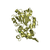
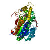
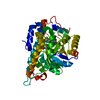
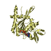
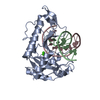
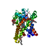
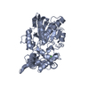
- PDB-5bu6: Structure of BpsB deaceylase domain from Bordetella bronchiseptica -
+ Open data

- Basic information
| Entry | Database: PDB / ID: 5bu6 | ||||||
|---|---|---|---|---|---|---|---|
| Title | Structure of BpsB deaceylase domain from Bordetella bronchiseptica | ||||||
 Components Components | BpsB (PgaB), Poly-beta-1,6-N-acetyl-D-glucosamine N-deacetylase | ||||||
 Keywords Keywords | HYDROLASE / deacetylase / family 4 carbohydrate esterase | ||||||
| Function / homology | Glycoside hydrolase/deacetylase / TIM Barrel / Alpha-Beta Barrel / Alpha Beta / NICKEL (II) ION / THIOCYANATE ION / :  Function and homology information Function and homology information | ||||||
| Biological species |  Bordetella bronchiseptica RB50 (bacteria) Bordetella bronchiseptica RB50 (bacteria) | ||||||
| Method |  X-RAY DIFFRACTION / SYNCHROTRON / MOLECULAR REPLACEMENT / Resolution: 1.951 Å X-RAY DIFFRACTION / SYNCHROTRON / MOLECULAR REPLACEMENT / Resolution: 1.951 Å | ||||||
 Authors Authors | Little, D.J. / Bamford, N.C. / Howell, P.L. | ||||||
| Funding support |  Canada, 1items Canada, 1items
| ||||||
 Citation Citation | Journal: J.Biol.Chem. / Year: 2015 Title: The Protein BpsB Is a Poly-beta-1,6-N-acetyl-d-glucosamine Deacetylase Required for Biofilm Formation in Bordetella bronchiseptica. Authors: Little, D.J. / Milek, S. / Bamford, N.C. / Ganguly, T. / DiFrancesco, B.R. / Nitz, M. / Deora, R. / Howell, P.L. | ||||||
| History |
|
- Structure visualization
| Structure viewer | Molecule: MolmilJmol/JSmol |
|---|
- Downloads & links
-Download
| PDBx/mmCIF format | 5bu6.cif.gz | 227.4 KB | Display | PDBx/mmCIF format |
|---|---|---|---|---|
| PDB format | pdb5bu6.ent.gz | 181.7 KB | Display | PDB format |
| PDBx/mmJSON format | 5bu6.json.gz | Tree view | PDBx/mmJSON format | |
| Others |  Other downloads Other downloads |
-Validation report
| Arichive directory | https://data.pdbj.org/pub/pdb/validation_reports/bu/5bu6ftp://data.pdbj.org/pub/pdb/validation_reports/bu/5bu6 | HTTPS FTP |
|---|
-Related structure data
| Related structure data | 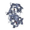 4f9dS S: Starting model for refinement |
|---|---|
| Similar structure data |


-Links
 PDBj
PDBj
- Assembly
| Deposited unit | 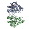
| ||||||||
|---|---|---|---|---|---|---|---|---|---|
| 1 | 
| ||||||||
| 2 | 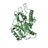
| ||||||||
| 3 | 
| ||||||||
| Unit cell |
|
-Components
| #1: Protein | Mass: 30928.957 Da / Num. of mol.: 2 / Fragment: UNP residues 35-307 / Mutation: C48S, K128A, K129A, D235H, E239H Source method: isolated from a genetically manipulated source Source: (gene. exp.) Bordetella bronchiseptica RB50 (bacteria)Strain: RB50 / Gene: hmsF, BN113_1689, BB1768 / Plasmid: pET28A / Production host: #2: Chemical | ChemComp-NI /   Mass: 58.693 Da / Num. of mol.: 5 / Source method: obtained synthetically / Formula: Ni Mass: 58.693 Da / Num. of mol.: 5 / Source method: obtained synthetically / Formula: Ni#3: Chemical | ChemComp-SCN /   Mass: 58.082 Da / Num. of mol.: 5 / Source method: obtained synthetically / Formula: CNS Mass: 58.082 Da / Num. of mol.: 5 / Source method: obtained synthetically / Formula: CNS#4: Chemical | ChemComp-EDO / |   Mass: 62.068 Da / Num. of mol.: 1 / Source method: obtained synthetically / Formula: C2H6O2 Mass: 62.068 Da / Num. of mol.: 1 / Source method: obtained synthetically / Formula: C2H6O2#5: Water | ChemComp-HOH / |  Mass: 18.015 Da / Num. of mol.: 125 / Source method: isolated from a natural source / Formula: H2O Mass: 18.015 Da / Num. of mol.: 125 / Source method: isolated from a natural source / Formula: H2O |
|---|
-Experimental details
-Experiment
| Experiment | Method: X-RAY DIFFRACTION / Number of used crystals: 1 |
|---|
- Sample preparation
| Crystal | Density Matthews: 2.2 Å3/Da / Density % sol: 44 % / Description: Small thin needles |
|---|---|
| Crystal grow | Temperature: 293 K / Method: vapor diffusion, hanging drop / Details: 30% PEG 2000 MME, 0.1 M potassium thiocyanate |
-Data collection
| Diffraction | Mean temperature: 100 K |
|---|---|
| Diffraction source | Source: SYNCHROTRON / Site: CLSI / Beamline: 08ID-1 / Wavelength: 0.979 Å |
| Detector | Type: RAYONIX MX-300 / Detector: CCD / Date: Jan 16, 2014 Details: DCM with cryo-cooled 1st crystal sagittally bent 2nd crystal followed by vertically focusing mirror |
| Radiation | Monochromator: double crystal Si(111) / Protocol: SINGLE WAVELENGTH / Monochromatic (M) / Laue (L): M / Scattering type: x-ray |
| Radiation wavelength | Wavelength: 0.979 Å / Relative weight: 1 |
| Reflection | Resolution: 1.95→50 Å / Num. obs: 39033 / % possible obs: 98.6 % / Observed criterion σ(I): -3 / Redundancy: 3.5 % / Biso Wilson estimate: 39.02 Å2 / Rmerge(I) obs: 0.07 / Net I/σ(I): 33 |
| Reflection shell | Resolution: 1.95→2.02 Å / Redundancy: 3.6 % / Rmerge(I) obs: 0.546 / Mean I/σ(I) obs: 3.2 / % possible all: 97.7 |
- Processing
| Software |
| |||||||||||||||||||||||||||||||||||||||||||||||||||||||||||||||||||||||||||||||||||||||||||||||||||||||||||||||||||||||||||||||||||||||||||||||||||||||||||||||||||||||||||||||||||||||||||||||||||||||||||||||||||||||||||||||||
|---|---|---|---|---|---|---|---|---|---|---|---|---|---|---|---|---|---|---|---|---|---|---|---|---|---|---|---|---|---|---|---|---|---|---|---|---|---|---|---|---|---|---|---|---|---|---|---|---|---|---|---|---|---|---|---|---|---|---|---|---|---|---|---|---|---|---|---|---|---|---|---|---|---|---|---|---|---|---|---|---|---|---|---|---|---|---|---|---|---|---|---|---|---|---|---|---|---|---|---|---|---|---|---|---|---|---|---|---|---|---|---|---|---|---|---|---|---|---|---|---|---|---|---|---|---|---|---|---|---|---|---|---|---|---|---|---|---|---|---|---|---|---|---|---|---|---|---|---|---|---|---|---|---|---|---|---|---|---|---|---|---|---|---|---|---|---|---|---|---|---|---|---|---|---|---|---|---|---|---|---|---|---|---|---|---|---|---|---|---|---|---|---|---|---|---|---|---|---|---|---|---|---|---|---|---|---|---|---|---|---|---|---|---|---|---|---|---|---|---|---|---|---|---|---|---|---|
| Refinement | Method to determine structure: MOLECULAR REPLACEMENT Starting model: 4F9D Resolution: 1.951→45.6 Å / SU ML: 0.22 / Cross valid method: FREE R-VALUE / σ(F): 1.35 / Phase error: 21.99 / Stereochemistry target values: ML
| |||||||||||||||||||||||||||||||||||||||||||||||||||||||||||||||||||||||||||||||||||||||||||||||||||||||||||||||||||||||||||||||||||||||||||||||||||||||||||||||||||||||||||||||||||||||||||||||||||||||||||||||||||||||||||||||||
| Solvent computation | Shrinkage radii: 0.9 Å / VDW probe radii: 1.11 Å / Solvent model: FLAT BULK SOLVENT MODEL | |||||||||||||||||||||||||||||||||||||||||||||||||||||||||||||||||||||||||||||||||||||||||||||||||||||||||||||||||||||||||||||||||||||||||||||||||||||||||||||||||||||||||||||||||||||||||||||||||||||||||||||||||||||||||||||||||
| Displacement parameters | Biso mean: 54 Å2 | |||||||||||||||||||||||||||||||||||||||||||||||||||||||||||||||||||||||||||||||||||||||||||||||||||||||||||||||||||||||||||||||||||||||||||||||||||||||||||||||||||||||||||||||||||||||||||||||||||||||||||||||||||||||||||||||||
| Refinement step | Cycle: LAST / Resolution: 1.951→45.6 Å
| |||||||||||||||||||||||||||||||||||||||||||||||||||||||||||||||||||||||||||||||||||||||||||||||||||||||||||||||||||||||||||||||||||||||||||||||||||||||||||||||||||||||||||||||||||||||||||||||||||||||||||||||||||||||||||||||||
| Refine LS restraints |
| |||||||||||||||||||||||||||||||||||||||||||||||||||||||||||||||||||||||||||||||||||||||||||||||||||||||||||||||||||||||||||||||||||||||||||||||||||||||||||||||||||||||||||||||||||||||||||||||||||||||||||||||||||||||||||||||||
| LS refinement shell |
| |||||||||||||||||||||||||||||||||||||||||||||||||||||||||||||||||||||||||||||||||||||||||||||||||||||||||||||||||||||||||||||||||||||||||||||||||||||||||||||||||||||||||||||||||||||||||||||||||||||||||||||||||||||||||||||||||
| Refinement TLS params. | Method: refined / Refine-ID: X-RAY DIFFRACTION
| |||||||||||||||||||||||||||||||||||||||||||||||||||||||||||||||||||||||||||||||||||||||||||||||||||||||||||||||||||||||||||||||||||||||||||||||||||||||||||||||||||||||||||||||||||||||||||||||||||||||||||||||||||||||||||||||||
| Refinement TLS group |
|
